
Fig. 6
- ID
- ZDB-IMAGE-170110-13
- Publication
- Xiao et al., 2016 - Chromatin-remodelling factor Brg1 regulates myocardial proliferation and regeneration in zebrafish
- All Figures
- Figures for Xiao et al., 2016
|
Fig. 6
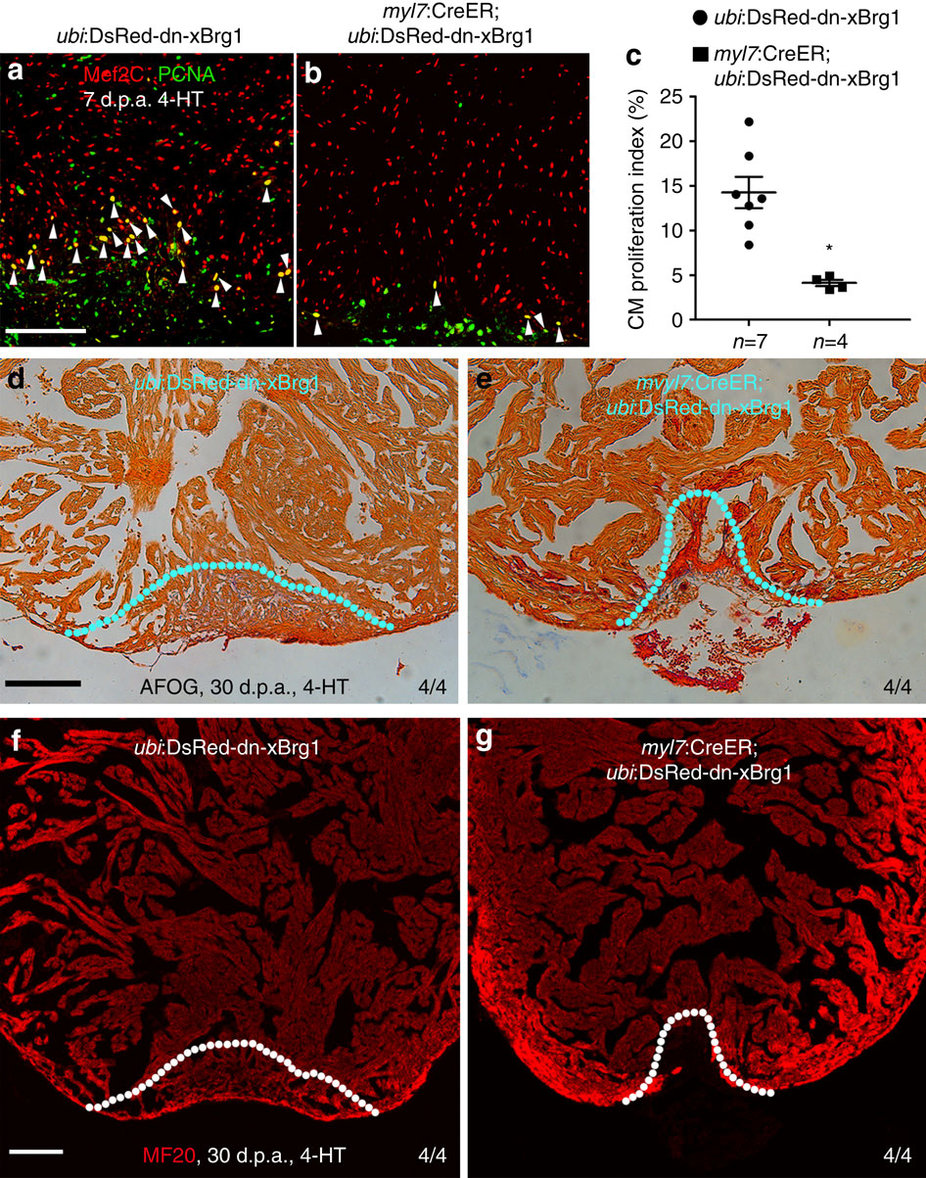
Myocardial-specific inhibition of Brg1 interferes heart regeneration.
(a–c) PCNA+/Mef2C+ proliferating cardiomyocytes decreased in Tg(myl7:CreER; ubi:DsRed-dn-xBrg1) transgenic hearts (b) compared with control Tg(ubi:DsRed-dn-xBrg1) transgenic hearts (a) at 7 d.p.a. Statistics of cardiomyocyte proliferation index is shown (*P<0.05; data presented are mean±s.e.m.; paired Student’s t-test) (c). White arrowheads point to PCNA+/Mef2C+ proliferating cardiomyocytes; n, the number of hearts analysed; ubi:DsRed-dn-xBrg1 stands for Tg(ubi:loxP-DsRed-STOP-loxP-dn-xBrg1); tamoxifen (4-HT) was applied at 3 days before injury. (d,e) AFOG staining revealed accumulated fibrin and fibrosis in Tg(myl7:CreER; ubi:DsRed-dn-xBrg1) transgenic hearts (e) compared with control Tg(ubi:DsRed-dn-xBrg1) transgenic hearts (d) at 30 d.p.a. (f,g) MF20 staining showed compromised myocardial regeneration in Tg(myl7:CreER; ubi:DsRed-dn-xBrg1) transgenic hearts (g) compared with control Tg(ubi:DsRed-dn-xBrg1) transgenic hearts (f) at 30 d.p.a. 4/4, all 4 hearts analysed showed the same phenotype. Scale bars, 100 μm.