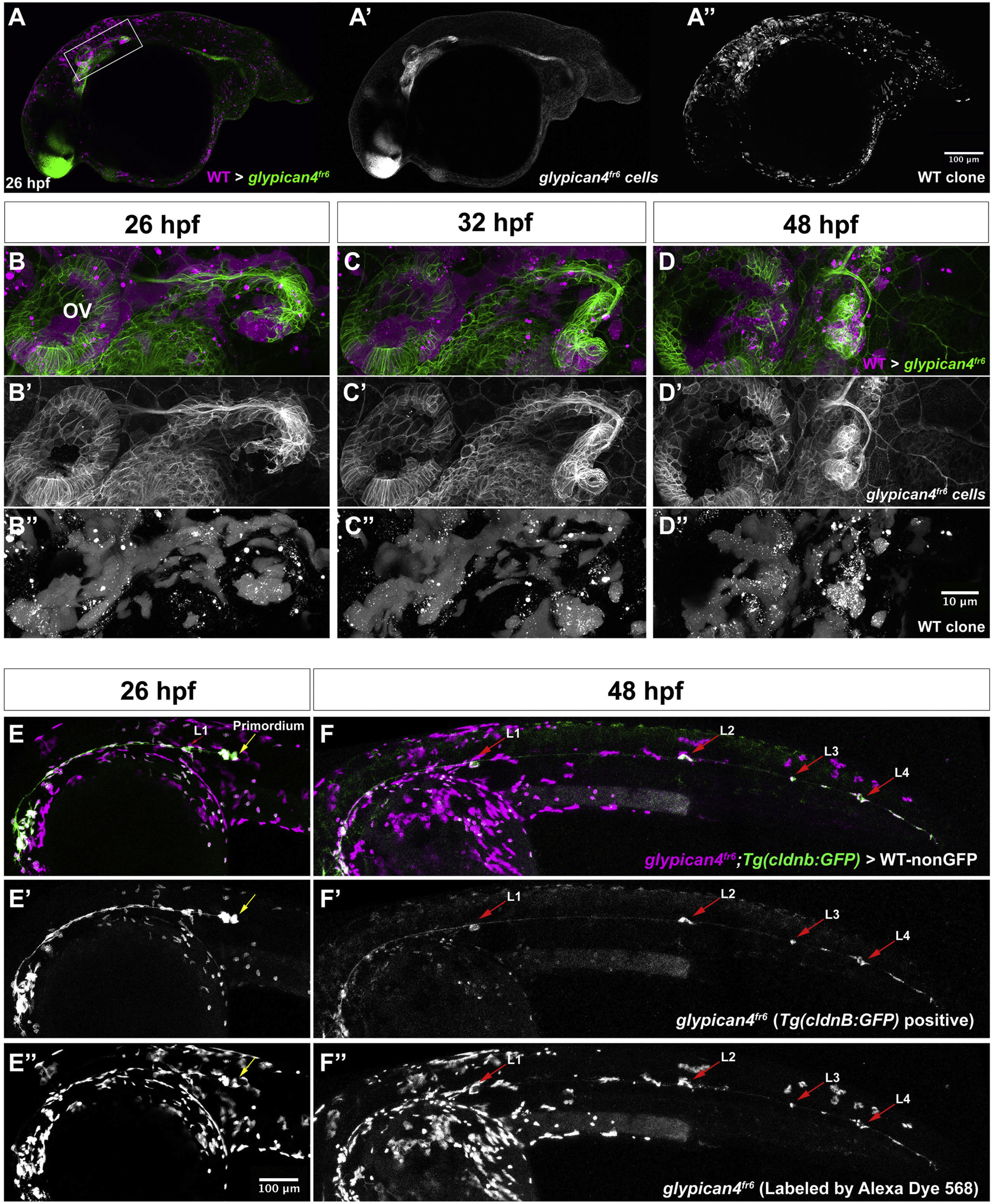
Fig. 7
glypican4fr6 mutant primordia display non-cell autonomous migration defects. (A, A′, A'') 26 hpf glypican4fr6;Tg(cldnb:lynGFP) mutant embryo into which wild type fluorescent Alexa-568 dye labeled cells were transplanted. Wild type cells are in magenta and glypican4fr6 cells are labeled green by cldnb:lynGFP. (A-A′) Wild type clones are found in the brain, skin, yolk, otic vesicle and primordium cells. (B) 40X magnification of the primordium clone boxed in (A) at 26 hpf showing magenta wild type cells in the otic vesicle (OV), lateral line ganglion and the primordium. Wild type primordium cells are not able to rescue the glypican4fr6 U-turning mutant phenotype and the primordium turns toward the ear. (C) Same primordium at 32 hpf. (D) By 48 hpf the mosaic primordium has stalled completely, irrespective of the number of wild type cells it contains demonstrating that glypican4 acts non cell-autonomously in the primordium. See Movie S3. (E-F”) glypican4fr6;Tg(cldnb:lynGFP) mutant cells (GFP+) labeled with fluorescent Alexa-568 dye were transplanted into a wild type embryo and primordium clones were screened at 26 hpf. (E-F) Shows a 10X image of the same embryo at 26 hpf and 48 hpf, respectively, in which the mutant primordium was able to complete migration and deposit neuromasts along its way to the tail tip. (B′, C′, D′, E′, F′) GFP+glypican4fr6 cells and (B”, C”, D”, E”, F”) red Alexa-568-labeled transplanted wild type cells.
Reprinted from Developmental Biology, 419(2), Venero Galanternik M., Lush, M.E., Piotrowski, T., Glypican4 Modulates Lateral Line Collective Cell Migration Non Cell-Autonomously, 321-335, Copyright (2016) with permission from Elsevier. Full text @ Dev. Biol.

