
IMAGE
Fig. 7
- ID
- ZDB-IMAGE-161219-24
- Publication
- Yang et al., 2016 - Deletion of Pr130 Interrupts Cardiac Development in Zebrafish
- All Figures
- Figures for Yang et al., 2016
Image
|
Figure Caption
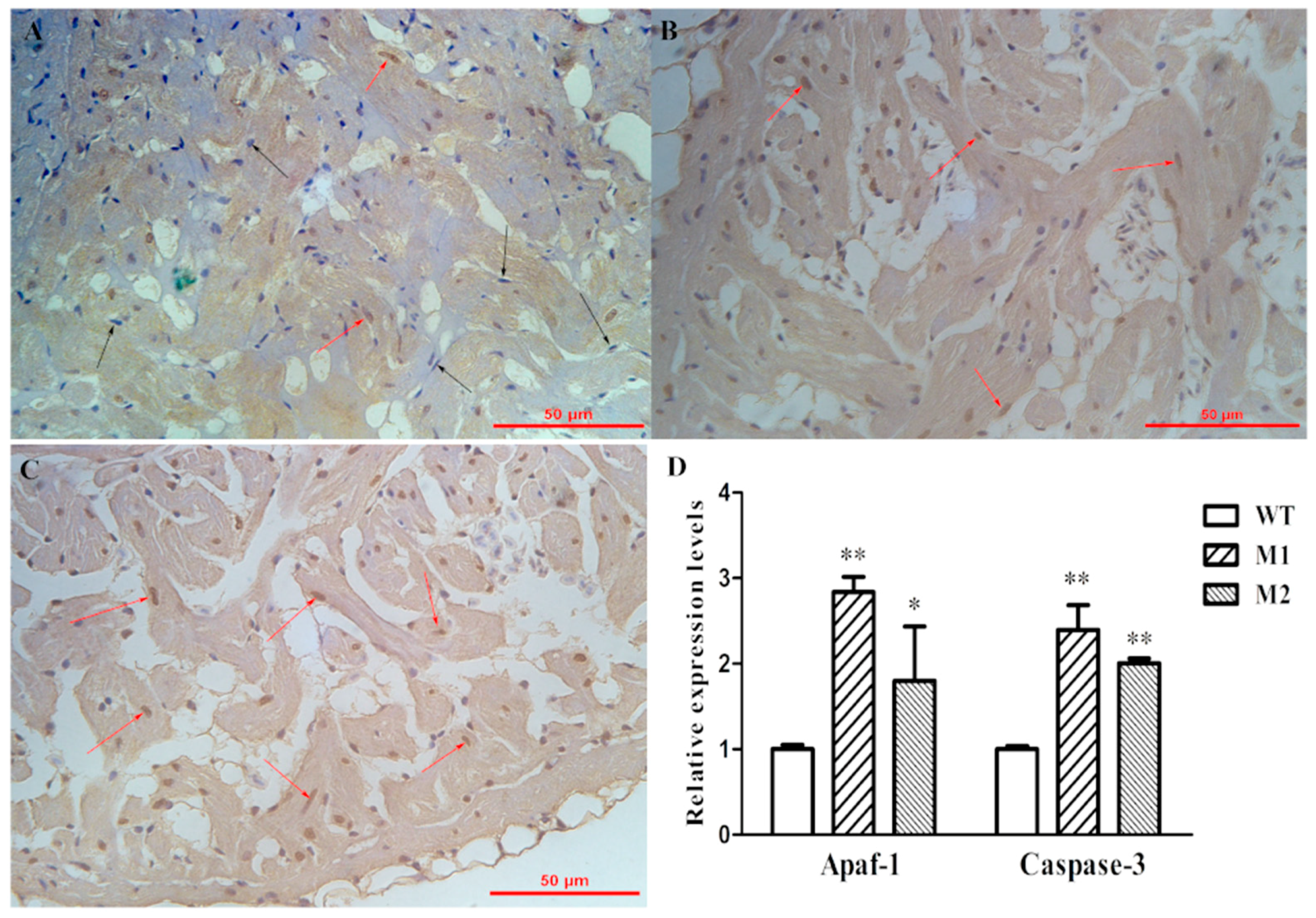
Fig. 7
Increased apoptosis was observed in pr130-/-. (A–C) Representative images of Tunel staining sections of adult heart tissue of WT (A), M1 (B), and M2 (C). Black arrows indicate normal cells (blue), red arrows indicate apoptosis cells (brown); (D) Expression levels of apoptosis-associated genes from adult heart tissues. Data represent the mean ± SD. The symbols * and ** in the bar chart represent significant differences (p < 0.05 or p < 0.01).
Figure Data
Acknowledgments
This image is the copyrighted work of the attributed author or publisher, and
ZFIN has permission only to display this image to its users.
Additional permissions should be obtained from the applicable author or publisher of the image.
Full text @ Int. J. Mol. Sci.