
|
Fig. 2
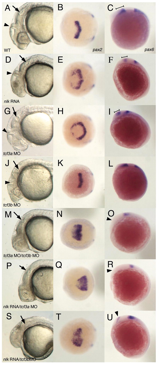
nlk RNA synergizes with tcf3a or tcf3b MOs. Comparison of wild-type (WT; A,B,C), nlk RNA (D,E,F), tcf3a MO (G,H,I), tcf3b MO (J,K,L), tcf3a/tcf3b MOs (M,N,O), nlk RNA/tcf3a MO (P,Q,R) and nlk RNA/tcf3b MO (S,T,U) embryos. nlk RNA (200 pg) was injected in conjunction with 1-2 ng of each of indicated morpholino. Control morpholino (see Materials and methods) was included to balance out the total amount of morpholino injected in each experiment. The first column shows lateral views of the head at 24 hours, with arrows indicating the otic vesicle and arrowheads marking the midbrain/hindbrain boundary (MHB). The middle column shows a dorsal view of embryos fixed at the 2 to 3 somite stage and stained with pax2 probe, whereas the last column shows lateral views of pax6 expression at 3 somites. In nlk RNA (D) and tcf3a MO (G) embryos, the eyes are missing, but embryos still develop a clear MHB, compared with wild type (A). pax2 expression is expanded in both nlk RNA (E) and tcf3a MO (H) embryos. pax6 expression in the diencephalon (marked by brackets in C,F,I) is slightly reduced in nlk RNA (F) and tcf3a MO (I) embryos relative to wild type (C). tcf3b MO embryos show a disorganization of the hindbrain (J), but still make eyes, and have normal pax2 and pax6 expression (K,L). Co-injection of tcf3a and tcf3b MOs results in more severe anterior truncations, with no visible MHB (M), a greatly expanded pax2 expression domain (N), and elimination of diencephalic pax6 (O, arrowhead). Co-injection of nlk RNA with either tcf3a MO (P,Q,R) or tcf3b MO (S,T,U) phenocopies the tcf3a/tcf3b MO phenotype.