Fig. 3
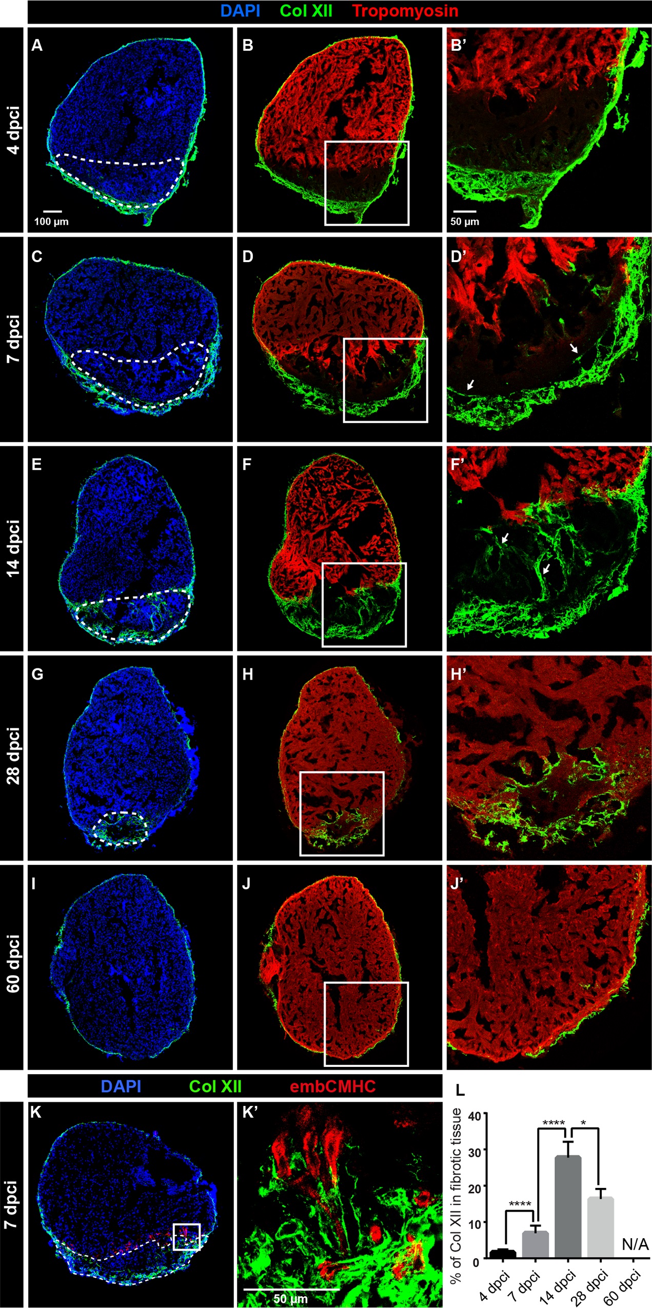
Collagen XII is transiently upregulated during heart regeneration in adult zebrafish.
(A-J) Immunostaining of transversal sections of ventricles at different time points after cryoinjury. The post-infarcted tissue is defined as a Tropomyosin-negative part of the ventricle (encircled with a dashed line). (A, B) At 4 dpci, the epicardial layer with Col XII becomes markedly thicker at the site of injury. N = 8. (C, D) At 7 dpci, the border zone of the remaining myocardium contains a few Col XII-positive fibrils (arrows). N = 8. (E, F) At 14 dpci, the post-cryoinjured tissue displays accumulation of Col XII-containing matrix. N = 7. (G, H) At 28 dpci, new cardiomyocytes replace the provisional tissue, leading to the progressive decrease of the post-cryoinjured matrix. Col XII persists in the leading edge of the regenerating myocardium. N = 5. (I, J) At 60 dpci, heart regeneration has been accomplished. The expression of Col XII is reminiscent of the original pattern of uninjured hearts. N = 8. (K) Immunofluorescence staining of ventricle sections using antibodies against Col XII (green) and embryonic cardiac myosin heavy chain (embCMHC, red). At 7 dpci, undifferentiated cardiomyocytes along the regenerating leading edge are associated with the Col XII-containing ECM. N = 6. (L) The percentage of Col XII-positive area within the fibrotic tissue of the post-cryoinjured myocardium without epicardial expression. N ≥ 5; **P < 0.01, **** P < 0.0001.

