
|
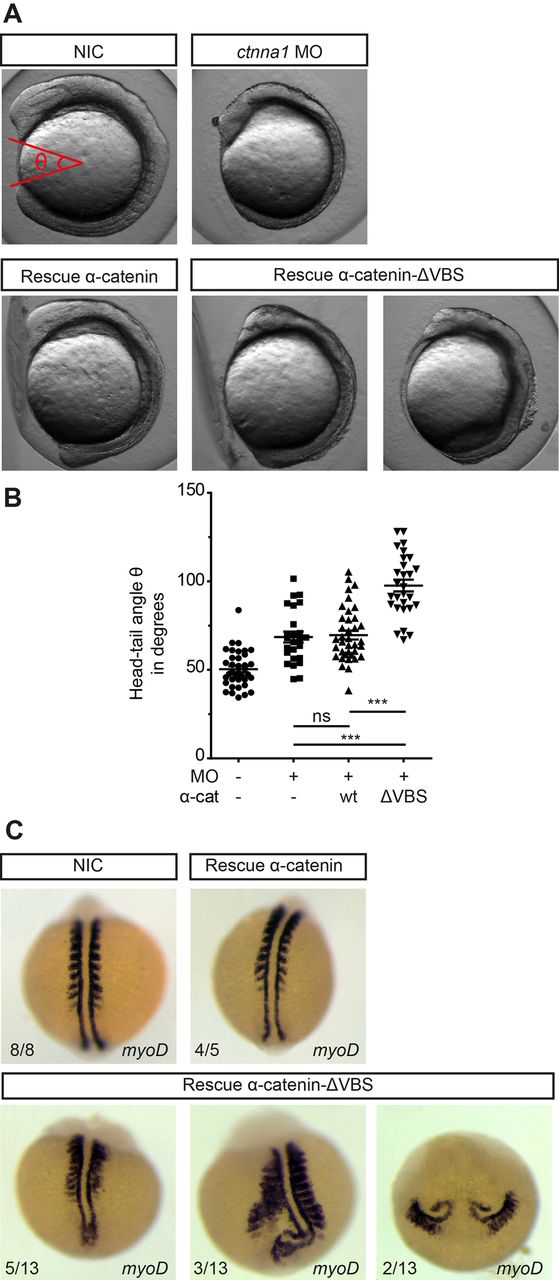
Fig. 5
Blocking vinculin recruitment to α-catenin induces convergent extension defects. (A) DIC images of non-injected embryos, embryos injected with 0.84 ng ctnna1 MO or ctnna1 MO-injected embryos rescued with either 250 pg α-catenin-GFP or 250 pg α-catenin-ΔVBS-GFP at the 5-7 somite stage (12 hpf). (B) Quantification of the angle between the anterior-most point in the head, and the posterior-most point in the tail of the embryos (shown in red in A). Datapoints represent single embryos from three independent experiments (n>25 embryos per condition). Data is represented as mean±s.e.m. One-way ANOVA following Tukey's multiple comparison test was used for comparisons among multiple groups, with ***P<0.0001. (C) Dorsal views of whole-mount in situ hybridization at 9-11 somite stage of non-injected embryos and ctnna1 MO-injected embryos rescued with either 250 pg α-catenin-GFP or 250 pg α-catenin-ΔVBS-GFP. MyoD marks the muscle precursors to visualize the somites.