
Fig. S2
- ID
- ZDB-IMAGE-161116-8
- Publication
- Rebman et al., 2016 - Cadherin-2 Is Required Cell Autonomously for Collective Migration of Facial Branchiomotor Neurons
- All Figures
- Figures for Rebman et al., 2016
|
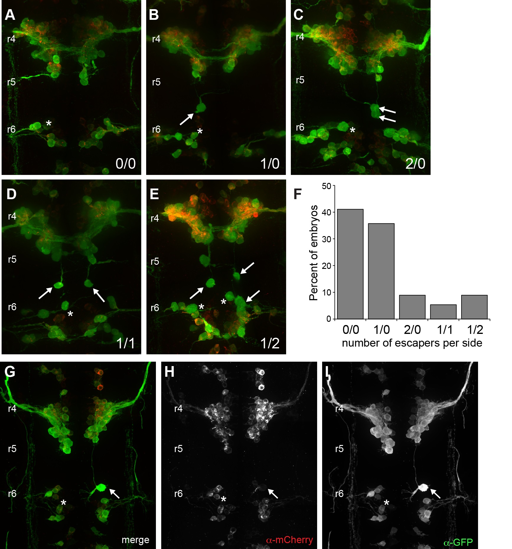
Fig. S2
Migration of ‘escaper’ neurons in homozygous Tg(isl1:cdh2ΔEC-mCherry)vc25 embryos.
(A-E) Confocal micrographs of dorsal views of homozygous Tg(isl1:GFP)/Tg(isl1:cdh2ΔEC-mCherry)vc25 embryos at 38 hpf. Embryos were labeled with α-GFP (green) and α-mCherry (red). Representative images of homozygous vc25Tg embryos that shows the majority of FBMNs fail to exit r4/r5 with or without a rare ‘escaper’ FBMN that migrates into r6 (arrows). (A) An embryo with no ‘escaper’ neurons present in r6 on either side of the midline (0/0). (B). An embryo with one ‘escaper’ neuron present in r6 on one side of the embryo, with no ‘escapers’ on the contralateral side (1/0). (C) An embryo with two ‘escaper’ FBMNs present in r6 on one side of the embryo and no ‘escapers’ on the contralateral side (2/0). (D) An embryo with one ‘escaper’ neuron present in r6 on both sides of the embryo (1/1). (E) An embryo with one ‘escaper’ neuron present in r6 on one side of the embryo and two ‘escaper’ FBMNs present on the contralateral side (1/2). (F) Histogram reflects the percentage of homozygous Tg(isl1:cdh2ΔEC-mCherry)vc25 embryos with each ‘escaper’ condition. (G-I) Confocal micrographs of immunostained embryos showing high magnification dorsal views of Tg(isl1:GFP)/Tg(isl1:cdh2ΔEC-mCherry)vc25 embryo at 38 hpf. White arrow shows ‘escaper’ neuron that expresses both isl1:GFP (green) and isl1:cdh2ΔEC-mCherry (red) transgenes, despite its presence in r6. White asterisk denotes r6-derived PLL efferent neurons, which differ from r4-derived FBMN populations.