
|
Fig. 3
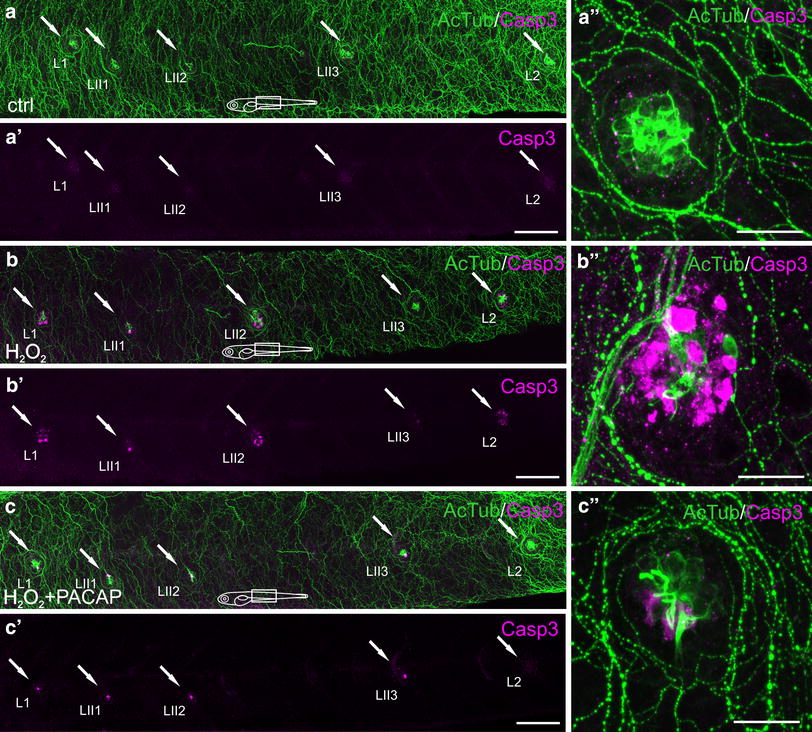
Immunohistochemical staining of the 5 dpf zebrafish neuromasts using antibodies against acetylated ±-tubulin (AcTub) (green) and caspase-3 (Casp3) (magenta). The visualization was accomplished using a Zeiss LSM-700 confocal microscope. The hair cells were stained with AcTub antibody and the apoptotic hair cells were marked with Casp3 antibody. a, b, c five trunk neuromasts (arrows from left to right: L1, LII1, LII2, LII3, L2, respectively) in the control animals (a), in the animals after 1 h 1.5 mM H2O2 exposure (b) and in the animals after 1 h 1.5 mM H2O2 + 100 nM PACAP-38 exposure preceded by 1 h preincubation with 100 nM PACAP-38 only (c) presented in a merged form with marked AcTub and Casp3 double-staining. a′-c′ Casp3 in hair cells of neuromasts L1, L II1, LII2, LII3, L2 (arrows) in the control animals (a′), in 1 h 1.5 mM H2O2-exposed group (b′) and in 1 h 1.5 mM H2O2 + 100 nM PACAP exposure + 1 h 100 nM PACAP preincubation group (c′) visualized without AcTub. a”–c′′ the presence of the apoptotic hair cells in the trunk neuromast L2 in the control animals (a′′), in the animals after 1 h 1.5 mM H2O2 exposure (b”) and in the animals after 1 h 1.5 mM H2O2 + 100 nM PACAP exposure preceded by 1 h preincubation with 100 nM PACAP only (c′′). The immunohistochemical staining is visualized in a merged form, where the AcTub and Casp3 double staining is distinctly marked. N = 15/group. Scale bars a, a′; b, b′; c, c′ 100 µm; a′′; b′′; c′′ 20 µm (Color figure online)