
Fig. 3
- ID
- ZDB-IMAGE-161107-22
- Genes
- Publication
- Marín-Juez et al., 2016 - Fast revascularization of the injured area is essential to support zebrafish heart regeneration
- All Figures
- Figures for Marín-Juez et al., 2016
|
Fig. 3
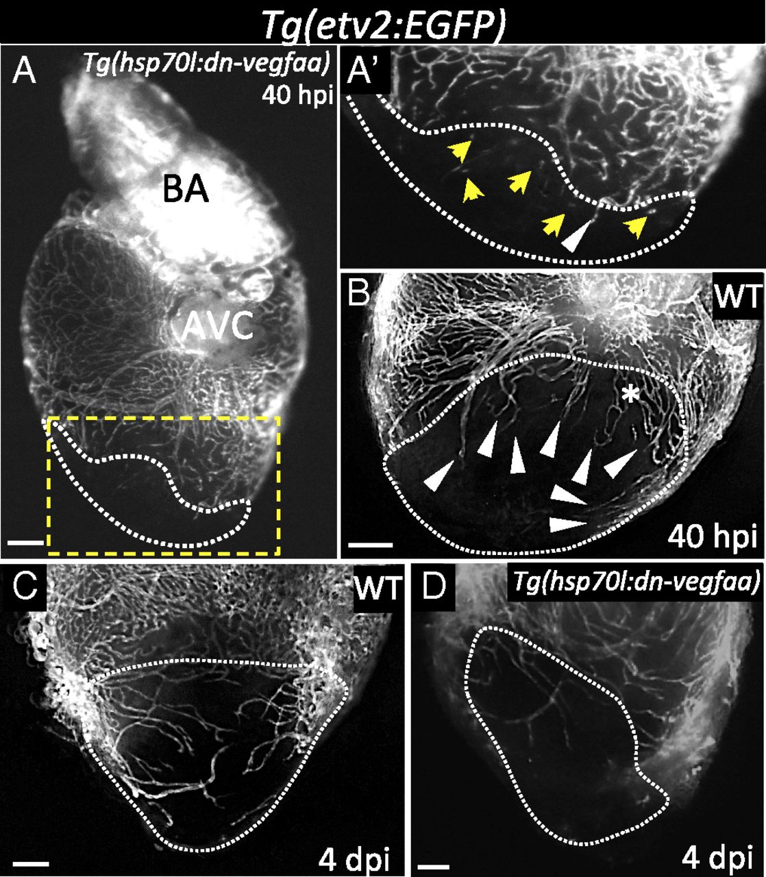
Induction of dn-vegfaa expression blocks revascularization of the damaged area. (A) Tg(hsp70l:dn-vegfaa);Tg(etv2:EGFP) ventricle at 40 hpi (n = 5). (A′) Inset shows high-magnification image of the injured area. (B) Tg(etv2:EGFP) WT ventricle at 40 hpi (n = 5). (C) Tg(etv2:EGFP) WT ventricle at 4 dpi (n = 5). (D) Tg(hsp70l:dn-vegfaa);Tg(etv2:EGFP) heart at 4 dpi (n = 3). White arrowheads point to coronary vessels sprouting into the injured area; yellow arrows point to disconnected endothelial cells; asterisk mark an area of coronary plexus. Dotted lines delineate the injured area. AVC, atrioventricular canal; BA, bulbus arteriosus. (Scale bar: 100 µm.)