
Fig. S1
- ID
- ZDB-IMAGE-161107-14
- Publication
- Marín-Juez et al., 2016 - Fast revascularization of the injured area is essential to support zebrafish heart regeneration
- All Figures
- Figures for Marín-Juez et al., 2016
|
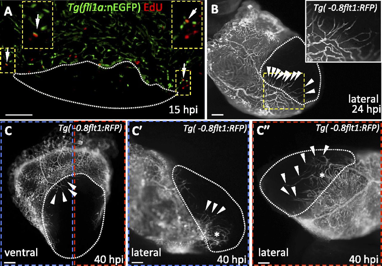
Fig. S1
Revascularization of the damaged area starts mainly from the dorsal half of the ventricle. (A) Section from a Tg(fli1a:nEGFP) ventricle stained for EdU incorporation. Arrows point to fli1a:nEGFP+/EdU+ nuclei in the ventricular wall. Insets show high-magnification images of fli1a:nEGFP+/EdU+ nuclei. (B) Lateral view of 24 hpi Tg(-0.8flt1:RFP) ventricle. Inset shows high-magnification image of vessels sprouting from preexisting coronaries. (C) Ventral view of 40 hpi Tg(-0.8flt1:RFP) ventricle. (C′ and C′′) Lateral views of 40 hpi Tg(-0.8flt1:RFP) ventricle. Arrowheads point to coronary vessels sprouting into the injured area; asterisk marks an area of coronary plexus (n = 5 ventricles examined). Dotted lines delineate the injured area. Arrowheads point to coronaries sprouting into the injured area. (Scale bars: 100 µm.)