
Fig. 1
- ID
- ZDB-IMAGE-161027-8
- Publication
- Cox et al., 2016 - Selenoprotein H is an essential regulator of redox homeostasis that cooperates with p53 in development and tumorigenesis
- All Figures
- Figures for Cox et al., 2016
|
Fig. 1
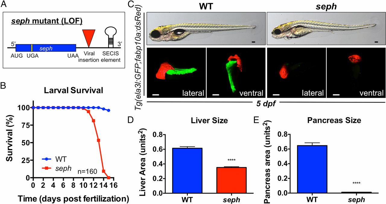
seph mutant zebrafish exhibit defects in organ development. (A) Schematic illustrating the nature of the seph mutant. (B) Survival of WT and homozygous seph mutant larvae over time. n = 160 larvae. (C) Morphological assessment and fluorescent imaging (lateral and ventral views) of WT and seph mutant larvae on a Tg(ela3l:GFP;fabp10a:dsRed) background by confocal tomography at 5 dpf. Liver volume: WT, 3.09e6 µm3; seph, 1.69e6 µm3. Pancreas volume: WT, 2.79e6 µm3; seph, undetectable. (Scale bar: 100 µm.) (D) Quantitative analysis of fluorescent liver area in WT and seph mutant larvae at 5 dpf. n = 16. ****P < 0.0001. (E) Quantitative analysis of fluorescent pancreas area in WT and seph mutant larvae at 5 dpf. n = 16. ****P < 0.0001.