
Fig. 1
- ID
- ZDB-IMAGE-161026-9
- Publication
- Deshwar et al., 2016 - Mespaa can potently induce cardiac fates in zebrafish
- All Figures
- Figures for Deshwar et al., 2016
|
Fig. 1
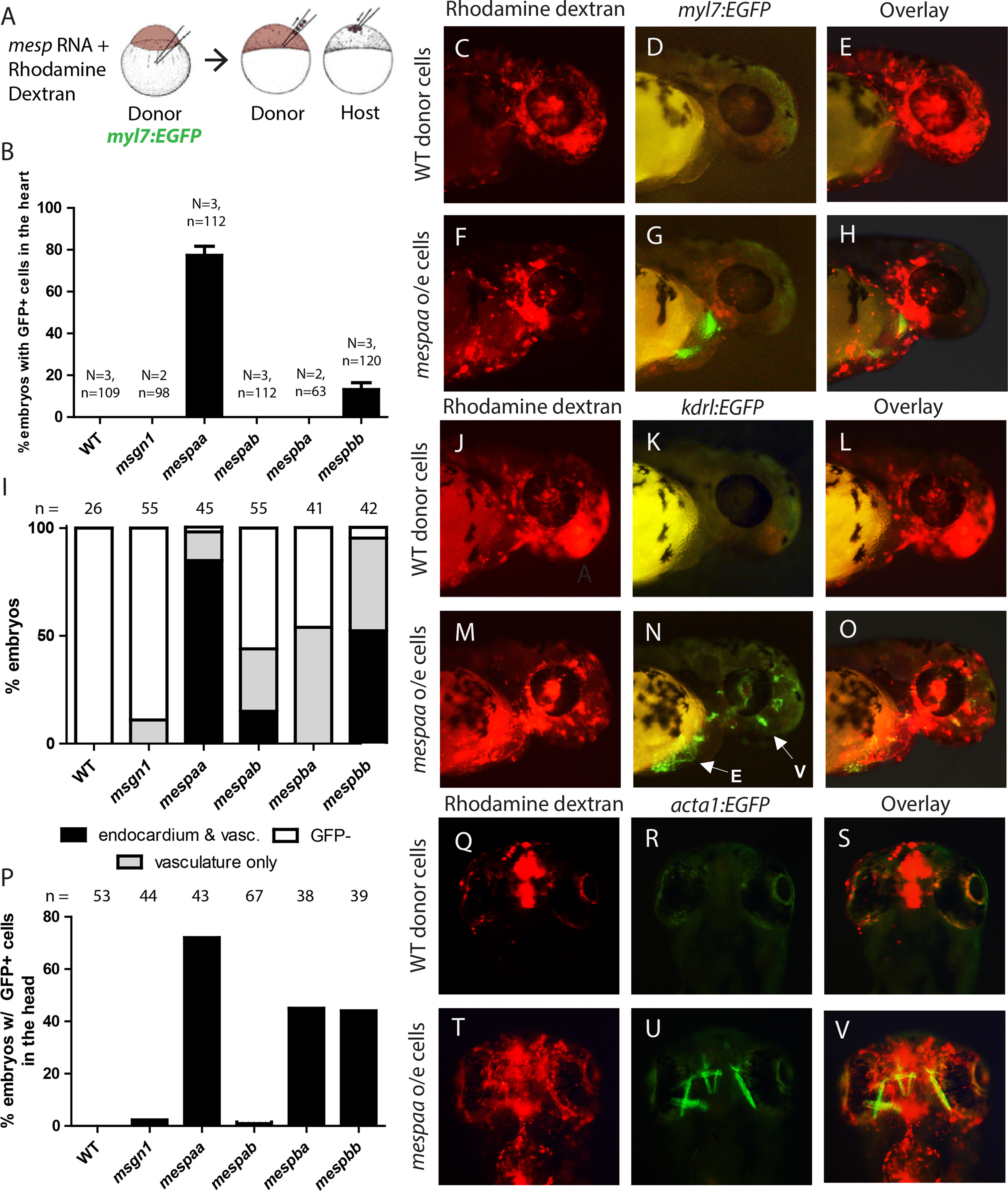
mesp over-expressing cells are capable of differentiating into myocardium, endocardium, vascular endothelium and facial muscle. (A) Schematic detailing transplantation experiments to assess the potential of mesp over-expressing cells. (B) Percent of host embryos with GFP-positive cells in the heart when different mesp family members were over-expressed in the transplant assay. (C-H) Host embryos at 48hpf generated from animal cap transplants of rhodamine dextran labeled myl7:EGFP donor cells of WT (C-E) or mespaa over-expressing cells (F-H). (I) Percent of host embryos with GFP-positive cells in the endocardium and vasculature or vasculature alone when different mesp family members were over-expressed in the transplant assay. (J-O) Host embryos at 48hpf generated from animal cap transplants of rhodamine dextran labeled kdrl:EGFP donor cells of WT (J-L) or mespaa over-expressing cells (M-O). E indicates endocardial cells, V designates vascular endothelial cells. (P) Percent of host embryos with GFP-positive cells in the head when different mesp family members were over-expressed. (Q-V) Host embryos at 48hpf generated from animal cap transplants of rhodamine dextran labeled acta1:EGFP donor cells of WT (Q-S) or mespaa over-expressing cells (T-V).
Reprinted from Developmental Biology, 418(1), Deshwar, A.R., Onderisin, J.C., Aleksandrova, A., Yuan, X., Burrows, J.T., Scott, I.C., Mespaa can potently induce cardiac fates in zebrafish, 17-27, Copyright (2016) with permission from Elsevier. Full text @ Dev. Biol.