
Fig. 4
- ID
- ZDB-IMAGE-161020-81
- Antibodies
- Publication
- Zhang et al., 2012 - AgRP and POMC neurons are hypophysiotropic and coordinately regulate multiple endocrine axes in a larval teleost
- All Figures
- Figures for Zhang et al., 2012
|
Fig. 4
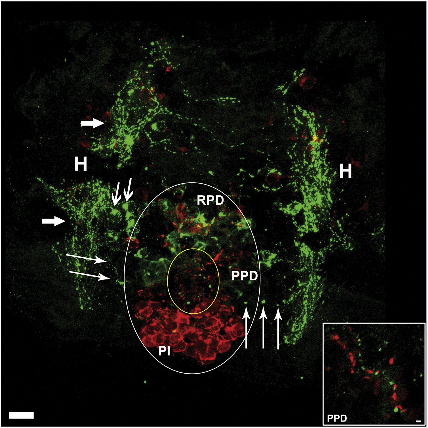
Hypothalamic AgRP- and α-MSH-Expressing Neurons Project to the Pituitary
Horizontal view of larval zebrafish brain at 5 dpf illustrating the pituitary and underlying hypothalamus. Large white oval approximates the extent of the pituitary, and small yellow oval indicates a zone within the proximal pars distalis containing dense α-MSH-immunoreactive (ir) fibers (red) and AgRP-ir fibers (green). Large arrows indicate hypothalamic AgRP-ir fiber bundles, medium arrows indicate hypothalamic AgRP-ir cell bodies, and thin arrows indicate AgRP-ir fibers projecting from hypothalamus into the pituitary. Inset is an enlargement from the PPD showing parallel AgRP-ir and α-MSH-ir neuronal fibers. PI, pars intermedia; PPD, proximal pars distalis; RPD, rostral pars distalis; H, hypothalamus. Scale bars: main image, 10 µm, inset = 1 µm.
Reprinted from Cell Metabolism, 15(2), Zhang, C., Forlano, P.M., and Cone, R.D., AgRP and POMC neurons are hypophysiotropic and coordinately regulate multiple endocrine axes in a larval teleost, 256-264, Copyright (2012) with permission from Elsevier. Full text @ Cell Metab.