
Fig. 3
- ID
- ZDB-IMAGE-161020-6
- Publication
- Granato et al., 1996 - Genes controlling and mediating locomotion behavior of the zebrafish embryo and larva
- All Figures
- Figures for Granato et al., 1996
|
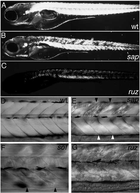
Fig. 3
Loss of birefringency and lesions in the somites become apparent in somite degeneration mutants. In sap (B) and ruz (C) mutants birefringency is decreased around 96 hours, compared to wild type (A), and lesions become visible in the somitic muscle tissue. (D) Lateral view of a wild-type larva around 96 hours; the somitic segments are separated by distinct boundries. (E,F) In sap and sof mutants degeneration affects a few individual somitic segments (black arrowheads indicate somite boundaries). The ventral part of the affected somite (E) appears normal (between white arrowheads). (G) In ruz mutants degeneration is affecting all somites.