
Image
|
Figure Caption
Fig. 3
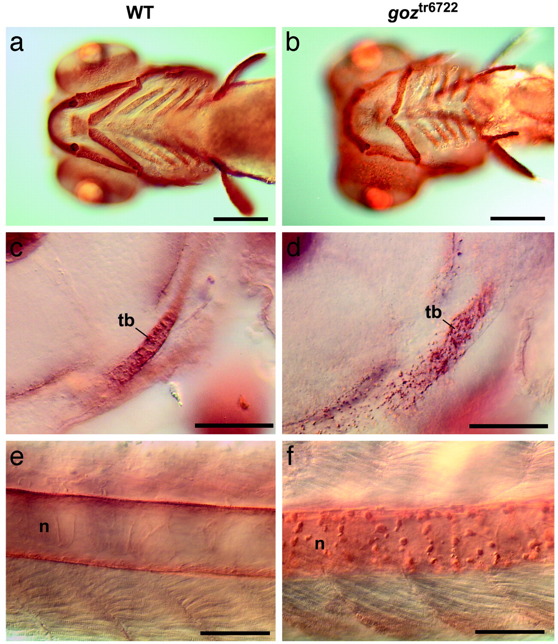
Col II defects in goz. Immunohistological staining of Col II in wild-type (WT) sibling larvae (a, c, and e) and goz larvae (b, d, and f) is shown. (a and b) Ventral view of the head 5 dpf. (c and d) Enlargement of the trabeculae (tb) of the neurocranium (lateral view) at 2 dpf. (e and f) Enlargement of the notochord (n) in the tail at 2 dpf. Cartilage matrix is homogeneously stained in sibling larvae (a, c, and e), whereas abnormal protein aggregates can be seen in the cartilage matrix and around the notochord in goz larvae (d and f). (Scale bars are 200 µm in a and b and 50 µm in c-f.)
Figure Data
Acknowledgments
This image is the copyrighted work of the attributed author or publisher, and
ZFIN has permission only to display this image to its users.
Additional permissions should be obtained from the applicable author or publisher of the image.
Full text @ Proc. Natl. Acad. Sci. USA