
|
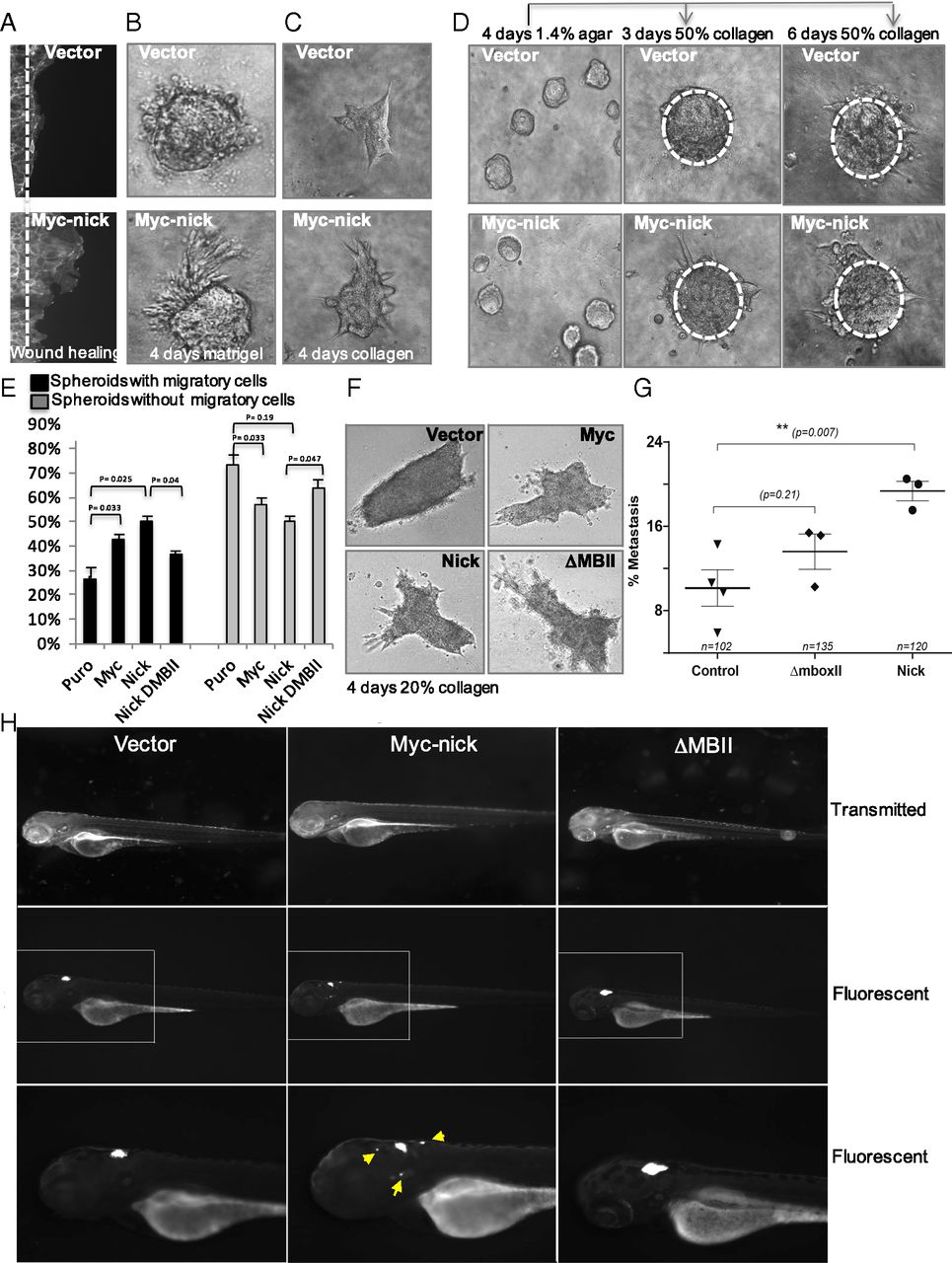
Fig. 2
Effect of MYC-nick on migration of colon cancer cells in 3D systems. (A) Scratch assay: DLD1 cells expressing MYC-nick or empty vector were grown on Cytoselect 24-Well plates for 48 h to confluency when the stopper was removed to allow migration. At 24 h later, cells were stained with phalloidin and photographed. (Magnification: 63×.) (B and C) The 3D culture assay: a total of 100 cells expressing MYC-nick or empty vector were trypsinized, and single cells were embedded in 50% (vol/vol) Matrigel (B) or collagen (C) matrix, grown for 3 d, and photographed. (D) Migration of colon cancer cells initially grown as spheroids for 3 d and then embedded in 50% (vol/vol) collagen for the indicated time points. (Magnification: B-D, 20×.) (E and F) Migration of DLD1 colon cancer cells expressing empty vector or MYC-nick in soft collagen. Cells were grown as spheroids over agar for 2 d and then were embedded in 20% (vol/vol) collagen. The percentage of spheroids displaying at least one migratory cell was calculated 24-h after seeding. Cultures were photographed 3 d after seeding (F). (Magnification: 20×.) (G and H). A total of 25-50 DLD1 cells expressing empty vector, MYC-nick, and MYC-nick lacking MYC box II (ΔMYC box II) were labeled with CellTracker Green, injected into the hindbrain of zebrafish embryos, and scored for migration (G) and photographed after 96 h.