
Fig. 4
- ID
- ZDB-IMAGE-161018-12
- Publication
- D'Orazi et al., 2016 - Mismatch of Synaptic Patterns between Neurons Produced in Regeneration and during Development of the Vertebrate Retina
- All Figures
- Figures for D'Orazi et al., 2016
|
Fig. 4
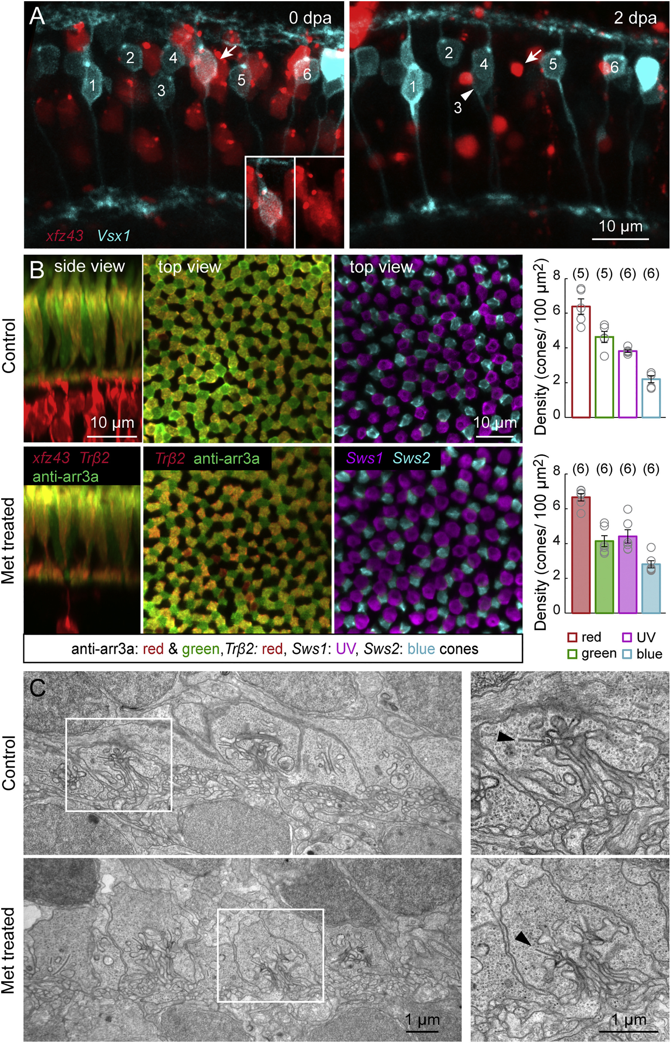
Nitroreductase-Induced Ablation of xfz43 BCs Does Not Damage Neighboring Cells
(A) Multiphoton time-lapse imaging of xfz43 BCs in the background of Tg(Vsx1:MCerulean) before and after cell ablation at 7 dpf. Larvae were imaged immediately prior to Met immersion, at 0 days post ablation (dpa), and after treatment, at 2 dpa. Vsx1 labels a population of BCs that includes a subset of xfz43 OFF BCs (arrow, inset). The somata of one of the Vsx1 BCs translocated between imaging sessions (arrowhead).
(B) Ablation of xfz43 BCs in the background of transgenically or immunostained cones. Red and green cones were visualized in xfz43 fish crossed with Tg(Trβ2:Tomato) larvae together with anti-arrestin3a immunostaining. UV and blue cones were visualized in xfz43 larvae crossed with Tg(Sws1:GFP; Sws2:mCherry). Maximum-intensity projections of confocal image stacks from 12 dpf (5 dpa) control or Met-treated retinas. Plots show the mean cell density of each cone type from Met-treated larvae at 5 dpa, and from age-matched control animals. Each open circle represents a single retina, with the number of retinas analyzed shown in parentheses. Error bars, ±SEM. Pairwise comparisons of the densities of each cone population between conditions showed no significant differences (p > 0.05, Wilcoxon-Mann-Whitney rank-sum test).
(C) Electron micrograph of the OPL in control and Met-treated retinas from 12 dpf xfz43 larvae. Insets show the ribbon synapses of single cone axon terminals. Arrowheads mark presynaptic ribbons.