
Fig. 2
- ID
- ZDB-IMAGE-161005-21
- Publication
- Ransom et al., 1996 - Characterization of zebrafish mutants with defects in embryonic hematopoiesis
- All Figures
- Figures for Ransom et al., 1996
|
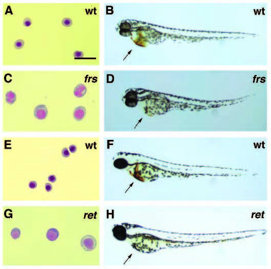
Fig. 2
Mutations in the genes frascati (frs) and retsina (ret) cause a reduction in the number of circulating blood cells. The remaining blood cells also have an abnormal cell morphology. (A,C,E,G) Isolated blood cells from wild-type and mutant larvae stained by the Wright- Giemsa method. (B,D,F,H) O-dianisidine staining for hemoglobin (arrows) in wild-type and mutant embryos. (A) Blood cells from 2-day old wild-type embryos have the normal morphology of erythroblasts. (B) Hemoglobin in red blood cells is strongly stained in wild-type embryos. (C) The circulating cells of 2-day old frstq223 mutant embryos are larger than normal erythroid cells and many have abnormally fragmented nuclei. (D) The number of blood cells in 2-day old frstq223 mutant embryos is greatly reduced which is reflected in the lower amount of hemoglobin. (E) Blood cells of 3-day old wild-type larva are small erythroblasts. (F) The 3-day old wild-type larva also has a large number of red blood cells producing hemoglobin. (G) The circulating cells of a 3-day old rettr217 mutant larva are larger and more basophilic than normal. (H) The 3-day old rettr217 mutant larva expresses very little hemoglobin, based on o-dianisidine staining. Scale bar (A,C,E,G) is equal to 20 µm.