
IMAGE
Fig. 4
- ID
- ZDB-IMAGE-161005-18
- Publication
- Weinstein et al., 1996 - Hematopoietic mutations in the zebrafish
- All Figures
- Figures for Weinstein et al., 1996
Image
|
Figure Caption
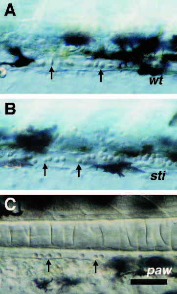
Fig. 4
Defects in erythrocyte morphogenesis in sti and paw mutants. DIC photomicroscopy of blood cells (arrows) in tail vessels of zebrafish embryos. (A) Phenotypically wild-type 2.25 dpf embryo from a cross between two stim232 heterozygotes; (B) mutant 2.25 dpf embryo from a cross between two stim232 heterozygotes; (C) mutant 2.25 dpf embryo from a cross between two pawm416 heterozygotes. Scale bar, 50 µm.
Figure Data
Acknowledgments
This image is the copyrighted work of the attributed author or publisher, and
ZFIN has permission only to display this image to its users.
Additional permissions should be obtained from the applicable author or publisher of the image.
Full text @ Development