
Fig. 3
- ID
- ZDB-IMAGE-161005-17
- Publication
- Weinstein et al., 1996 - Hematopoietic mutations in the zebrafish
- All Figures
- Figures for Weinstein et al., 1996
|
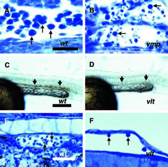
Fig. 3
Blood cells are not properly generated during early stages of hematopoiesis in vmp and vlt mutants. Embryos were obtained from crosses between two vmpm62 heterozygotes (A,B) or two vltm651 heterozygotes (C-F). (A,C,E) Phenotypically wild-type embryos. (B,D,F) Mutant siblings. (A,B) Wright-Giemsa stained longitudinal sections from the tails of 27 somite stage (approximately 22.5 hpf) mutant (B) and unaffected sibling (A) embryos. Erythroblasts are seen in both embryos (arrows), but there are many fewer are present in mutants. (C,D) Whole DAF stained 28 somite stage (23 hpf) phenotypically wild-type (C) and mutant (D) embryos. DAF staining is seen in the IMC of wild-type but not mutant embryos (arrows). (E,F) Wright-Giemsa stained longitudinal sections of 2.25 dpf phenotypically wild-type (E) and mutant (F) embryos. Normal blood cells are seen in wild-type embryos, but only abnormal, large cells are seen rarely in mutants (arrows). Scale bars 25 µm (A,B), 200 µm (C,D) and 50 µm (E,F).