
Fig. 2
- ID
- ZDB-IMAGE-160929-6
- Publication
- Jung et al., 2016 - Impaired Lymphocytes Development and Xenotransplantation of Gastrointestinal Tumor Cells in Prkdc-Null SCID Zebrafish Model
- All Figures
- Figures for Jung et al., 2016
|
Fig. 2
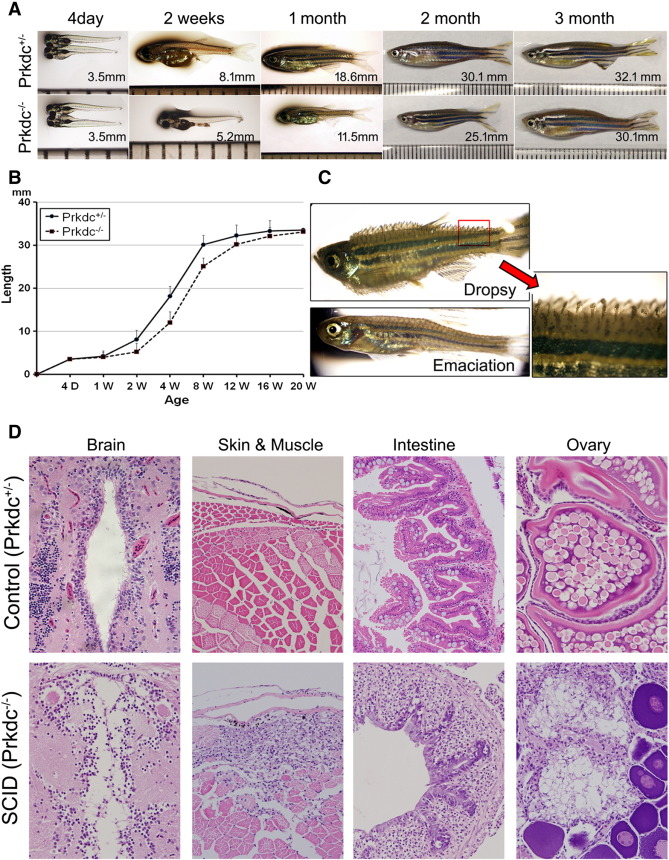
Growth phenotypes and microscopic observations of the Prkdc-null SCID zebrafish. (A and B) Growth retardation. The SCID zebrafish showed a retarded growth in length compared with the age-matched wild-type controls until 3 months. (C) Infectious manifestation. The immune-deficient SCID zebrafish often succumbed to infection. The emaciated zebrafish died within 7 days, whereas the "dropsy" zebrafish died within a couple of days. Red arrow shows a magnified view of the dropsy zebrafish fin. (D) Microscopic findings of the infection and inflammation occurred in the internal organs. The zebrafish with "dropsy" phenotype revealed that the disseminated infection occurred in the internal organs including brain, intestine, and ovary, suggesting septicemia. The emaciated zebrafish showed a predominant muscle necrosis with inflammation.