
Fig. 6
- ID
- ZDB-IMAGE-160929-11
- Publication
- Jung et al., 2016 - Impaired Lymphocytes Development and Xenotransplantation of Gastrointestinal Tumor Cells in Prkdc-Null SCID Zebrafish Model
- All Figures
- Figures for Jung et al., 2016
|
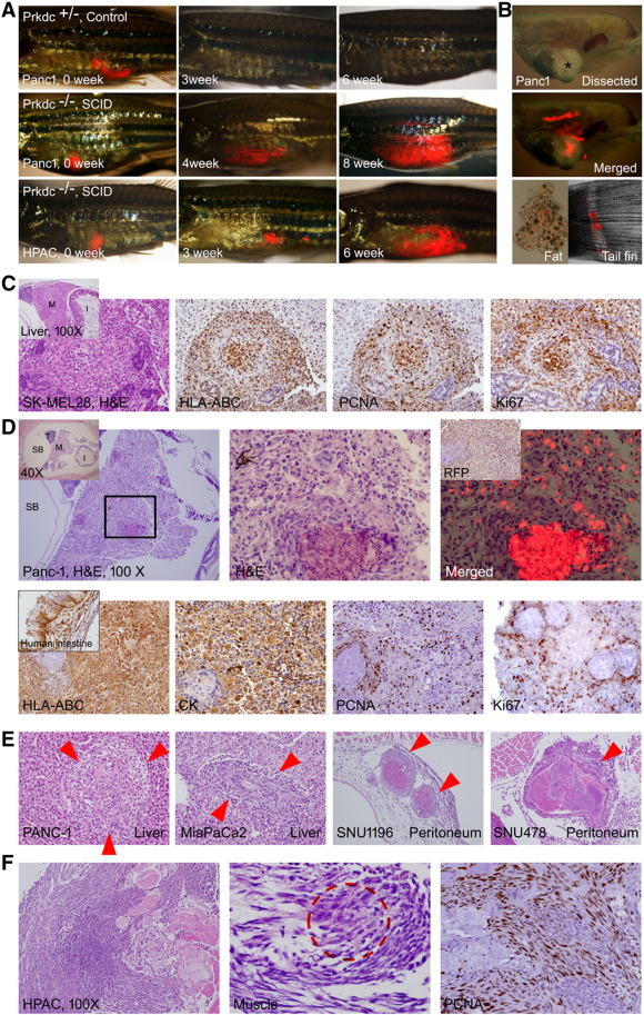
Fig. 6
Xenograft of tumor cells. Various tumor cells (5 × 10⁴) expressing TurboRFP were injected into intraperitoneal space by using a Hamilton syringe. If not indicated, all H&E and IHC images are 400×. (A) Gross images. The engraftment allows repeated observation of the growing tumors by visualization of transcutaneous RFP. Tumor engraftment was never formed in Prkdc+/-. (B) Dissected images. Bright field (top, asterisk shows a mass formed by the growing tumor) and merged RFP (middle) images showing an engrafted tumor with RFP expression. Dissected fat tissue (bottom left) showed scattered RFP-expressing tumor cells. Metastatic tumor cells (bottom right) at tail fin are also noted. (C) A mass formed in the liver in SK-MEL28 injected zebrafish. Inset is a low-power view showing mass (M) in the liver. HLA-ABC positivity of the tumor cells indicates that the mass originated from injected human melanoma cells. Many of the tumor cells are positive for PCNA and Ki-67, suggesting active proliferation. (D) A discrete mass formed in the Panc-1–injected zebrafish. Inset in the left is a low-power view indicating testis (T), swim bladder (SB), intestine (I), and mass (M). Enlarged view of the boxed area is shown in the middle. Merged TurboRFP and H&E image is shown on the right. Inset in the right is IHC for RFP. Lower column images are findings of IHC stain. Xenografted tumor cells are positive for HLA-ABC, for pan-cytokeratin (Pan-CK), and also for PCNA and Ki-67 frequently. (E) The tumors formed in liver and peritoneal cavity. (F) The xenografted HPAC cells showing the invasive growth, which results in destruction of muscle (M) and skin. The fibroblast with occasional tumor cells (dotted red circle shown in the middle). The proliferating fibroblasts are highly positive to PCNA.