
Fig. 9
- ID
- ZDB-IMAGE-160928-17
- Genes
- Publication
- Sarmah et al., 2016 - Embryonic Ethanol Exposure Dysregulates BMP and Notch Signaling, Leading to Persistent Atrio-Ventricular Valve Defects in Zebrafish
- All Figures
- Figures for Sarmah et al., 2016
|
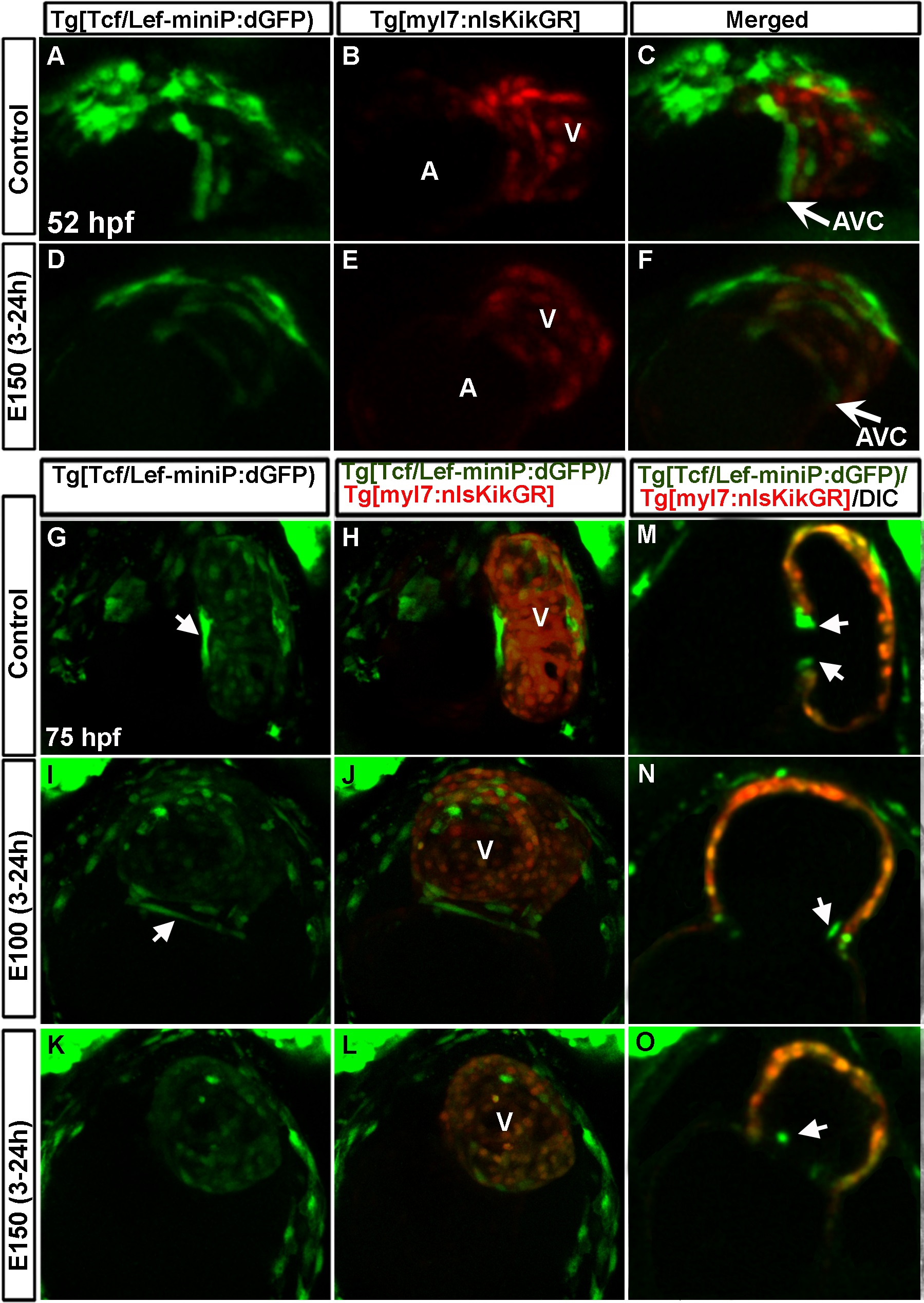
Fig. 9
Wnt activity was reduced in heart during AVC differentiation due to embryonic ethanol exposure.
(A-F) Tg(Tcf/Lef-miniP:dGFP);Tg(myl7:nlsKikGR) double transgenic zebrafish showed two rows of cardiomyocytes (red cells) at the AVC expressing GFP (Wnt active) in control embryos (A-C); ethanol treated embryos showed weak GFP expressing cardiomyocytes that were not restricted at the AVC (D-F) at 52 hpf. (G-L) Tg(Tcf/Lef-miniP:dGFP);Tg(myl7:nlsKikGR) double transgenic zebrafish showed long cardiomyocytes with strong GFP signal at the AVC and faint GFP signal in the ventricular cardiomyocytes in control embryos (G, H); ethanol exposed embryos showed weak GFP labeling at the AVC as compared to control (I-L). Arrows: Long cardiomyocytes with strong GFP signal. (M-O) Optical section of the heart of 75 hpf embryos showed GFP expression in the endocardial cells (arrowheads) at the AVC in the control embryo (M) and in ethanol treated embryos (N, O). A: Atrium, V: ventricle.