
Fig. 6
- ID
- ZDB-IMAGE-160928-14
- Genes
- Publication
- Sarmah et al., 2016 - Embryonic Ethanol Exposure Dysregulates BMP and Notch Signaling, Leading to Persistent Atrio-Ventricular Valve Defects in Zebrafish
- All Figures
- Figures for Sarmah et al., 2016
|
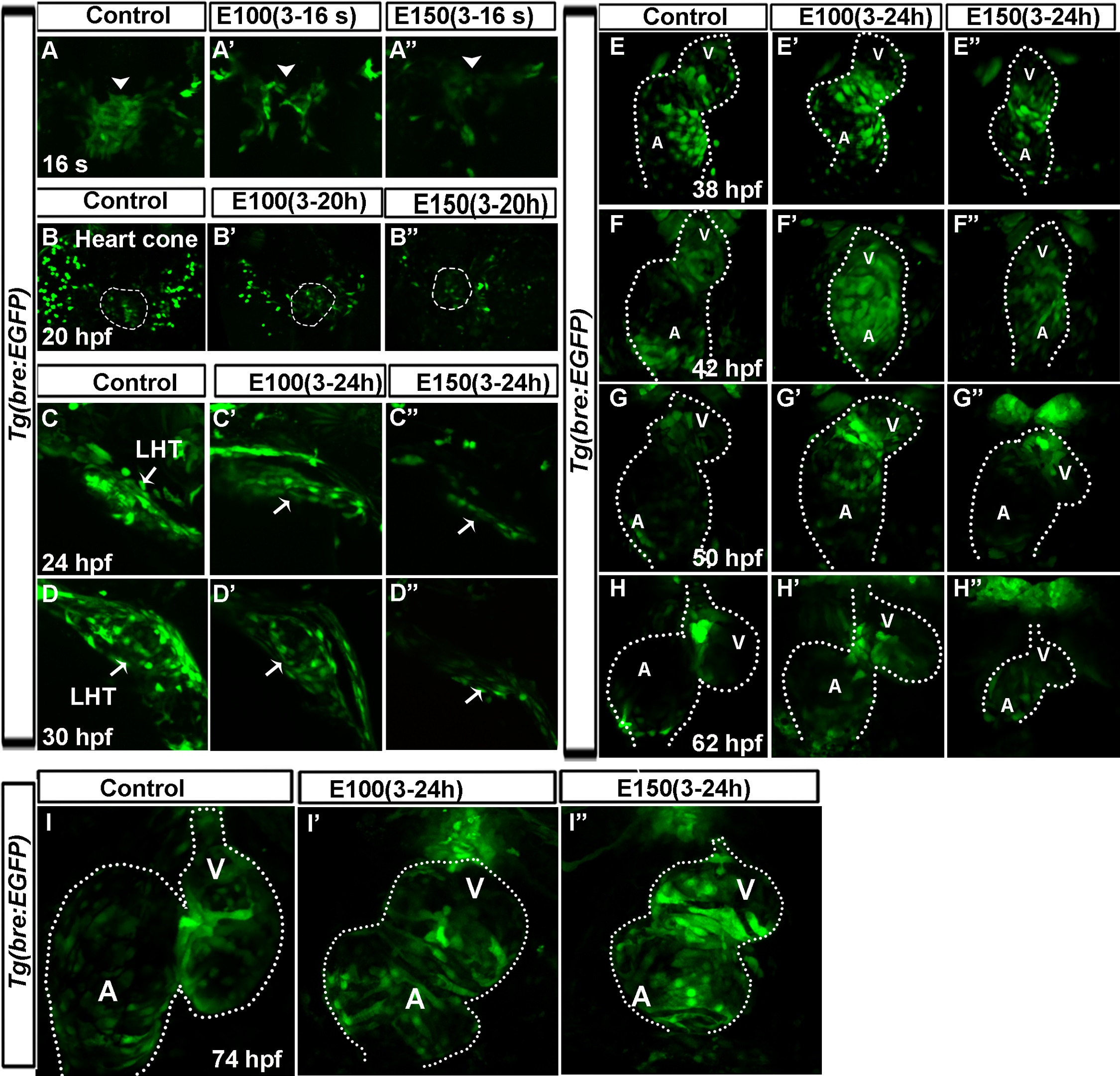
Fig. 6
Ethanol exposure reduced Bmp activity during heart tube morphogenesis, and later during valvulogenesis, those embryos lost regionalization of Bmp activity at the AVC.
(A-B′′) Tg(bre:EGFP) embryos showed reduced GFP label in the cardiac primordia in ethanol exposed embryos (A′-B′′) relative to control (A, B) at 16 somite (16s; 17 hpf; A-A′′) and heart cone stage (20 hpf; B-B′′). Arrowheads: cardiomyocytes; dotted line demarcates the heart cone. (C-D′′) Reduced GFP signaling was observed in the linear heart tube (LHT) of ethanol exposed embryos at 24 (C′, C′′) and 30 hpf (D′, D′′) compared to control (C, D). (E-I′′) Examination of Bmp activity from 38-74 hpf showed progressively restricted Bmp activity in the chamber cardiomyocytes and strong activity at the AVC in control embryos (E-I); ethanol treated embryos showed lack of restriction of Bmp activity at the AVC (E′-I′′) and occasional weak BMP activity in the heart (H′′). Dotted line demarcates the heart. A: Atrium, V: Ventricle.