
Fig. 4
- ID
- ZDB-IMAGE-160928-12
- Genes
- Publication
- Sarmah et al., 2016 - Embryonic Ethanol Exposure Dysregulates BMP and Notch Signaling, Leading to Persistent Atrio-Ventricular Valve Defects in Zebrafish
- All Figures
- Figures for Sarmah et al., 2016
|
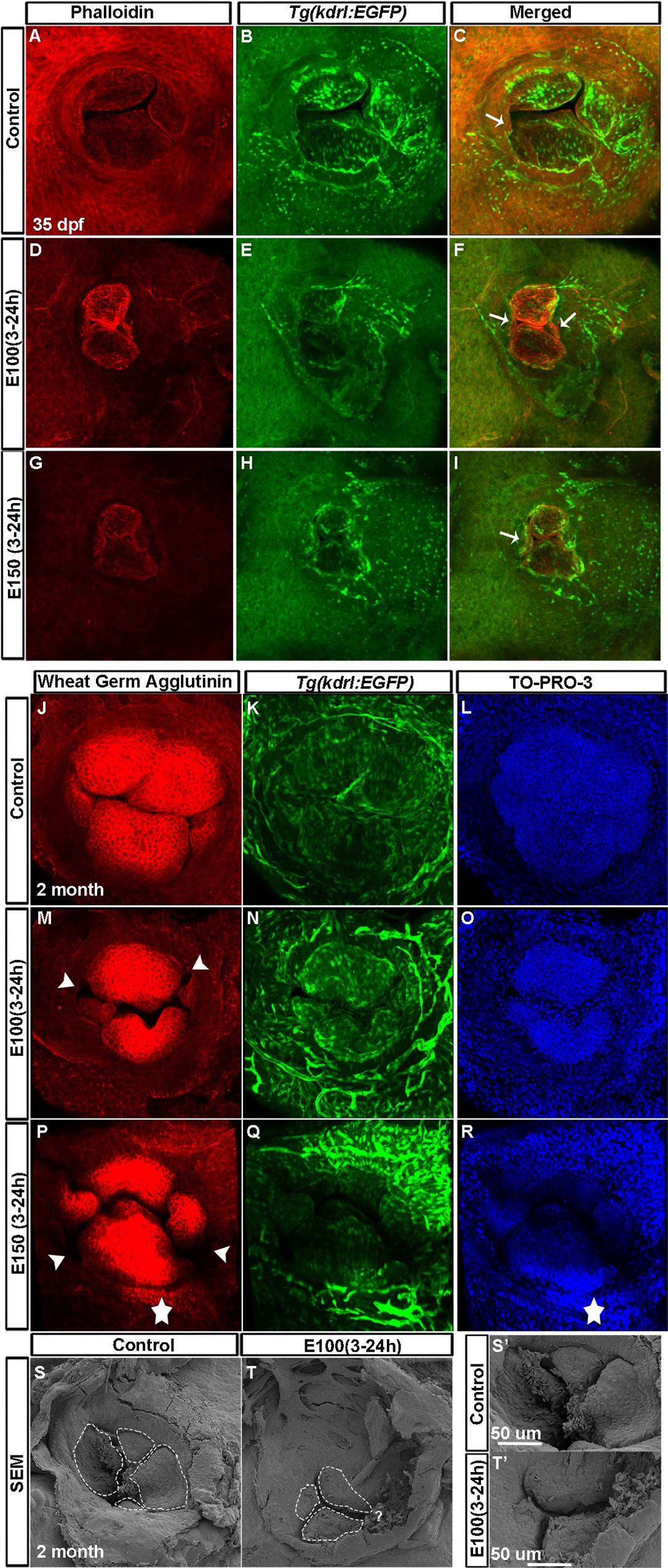
Fig. 4
Juvenile fish exposed to ethanol during embryonic development exhibited defective valves.
(A-I) Phalloidin stained Tg(kdrl:EGFP) fish showed two bigger valve cusps at the superior and inferior position, and two smaller leaflets oriented left and right of the AV orifice in control fish at 35 dpf (A-C); ethanol-exposed fish showed four smaller and irregular-shaped valve cusps (D-I). Arrows: smaller cusps. (J-R) WGA stained 2 months old Tg(kdrl:EGFP) fish exposed to ethanol during embryonic development showed four well-organized nicely-shaped valve cusps in control fish (J-L) and smaller, malformed valves in ethanol exposed fish (M-R). Arrowheads point to the holes between valve cusps; star demarcates the region strongly labeled by WGA at the AV orifice. (S-T′) Scanning electron microscopy of two months old fish showed four valve cusps in control fish (S-S′) and three smaller valve leaflets in ethanol treated fish (T, T′). S′ and T′: high magnification views of S and T; "?" point to the region of the possible fourth cusp.