
Fig. 3
- ID
- ZDB-IMAGE-160928-11
- Genes
- Publication
- Sarmah et al., 2016 - Embryonic Ethanol Exposure Dysregulates BMP and Notch Signaling, Leading to Persistent Atrio-Ventricular Valve Defects in Zebrafish
- All Figures
- Figures for Sarmah et al., 2016
|
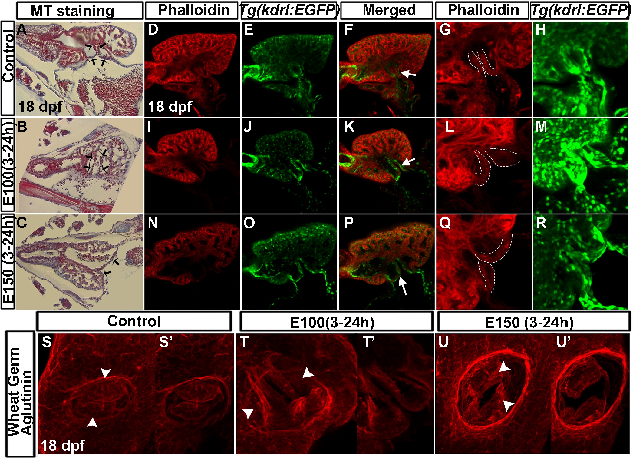
Fig. 3
Late-staged zebrafish larvae exposed to ethanol during embryogenesis exhibited defective valve leaflets.
(A-C) Masson′s trichrome stained coronal heart sections of 18 dpf larvae showed short and curved valve leaflets in control larvae (A), and longer and straight valve leaflets in larvae exposed to ethanol during embryogenesis (B, C). Black arrows: valve leaflets. (D-R) Single optical sagittal section of the heart of phalloidin stained Tg(kdrl:EGFP) larvae showed short AV valve leaflets in control larvae (D-F) and long valve leaflets in ethanol exposed larvae (I-K, N-P). White arrows: valve leaflets. 3D-rendering of the confocal sections showed narrow valve leaflets in the control (G, H) and thickened valve leaflets in ethanol exposed larvae (L, M, Q, R). White dotted lines demarcate valve leaflets. (S-U′) 3D-rendering of the optical sections at the AV junction of WGA labeled larvae showed two valve cusps in control larvae (S, S′) and deformed valve cusps in ethanol-exposed larvae (T-U′); S (54 µM thick), T (79.38 µM thick), U: 3D (81.9 µM thick) show the rendering of total number of Z sections. S′, T′, U′: 3D-rendering of same number of Z sections (25.2 µM thick). MS: Masson′s trichrome, Arrowhead: valve cusps.