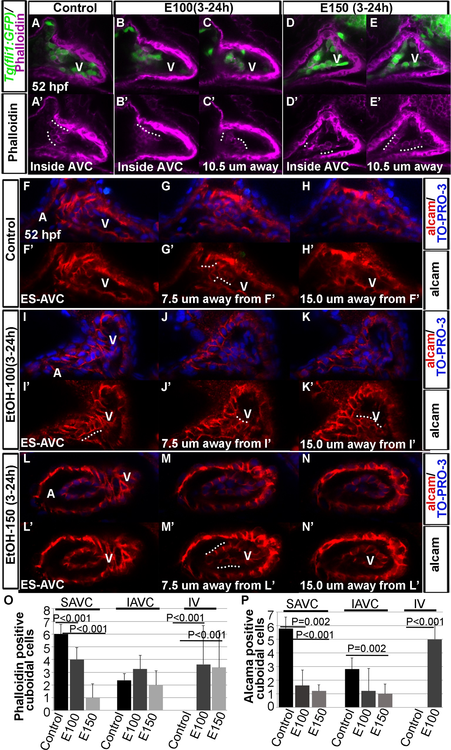
Fig. 2
Ethanol-exposure reduced precursors of valves at the AVC and produced ectopic valve-like cells in the ventricular endocardial lining.
(A-E′) Confocal sections of phalloidin stained Tg(fli1:EGFP) embryos showed differentiated cuboidal valve precursors inside AVC in control embryos (A, A′); E100 ethanol treated embryos showed phalloidin stained cuboidal cells inside AVC (B, B′) and inside the ventricle (10.5 µM away from the section B; C, C′); E150 ethanol treated embryos showed fewer phalloidin stained cuboidal cells at the AVC (D, D′) but more cells inside the ventricle (10.5 µM away from the section D; E, E′). White dotted lines show the rows of cuboidal cells. (F-N′) Confocal sections of anti-Alcama antibody stained embryos showing Alcama expression in the endocardial cells at the surface of the endocardium at AVC (F, F′, I, I′, L, L′), 7.5 µM (G, G′, J, J′, M, M′) and 15.0 µM away from surface endocardial cell sections (H, H′, K, K′, N, N′) in control and ethanol-exposed embryos. Control embryos showed Alcama positive cuboidal cells at 7.5 µM away from the surface AVC endocardium (G, G′); ethanol-exposed embryos showed Alcama positive cuboidal cells at the surface of AVC endocardium (I, I′) as well as 7.5 µM and 15 µM away from the AVC (J-K′, M-M′). White dotted lines show rows of cuboidal cells. A: Atrium, V: Ventricle, ES-AVC: Endocardial cells at the surface of AVC. (O, P) Graphs depict the number of phalloidin positive (O) and Alcama positive (P) cuboidal cells at the superior (SAVC) and inferior (IAVC) aspects of AVC, and inside the ventricle (IV) in control and ethanol-treated embryos. Statistically significant P values are shown in the graphs.

