
|
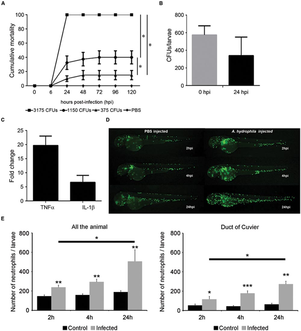
Fig. 3
Infection model by microinjection in the duct of Cuvier. (A) Cumulative mortality in larvae injected with various doses of bacteria (3175 CFU/nL, 1150 CFU/nL, and 375 CFU/nL). Graph shows a representative experiment of three independent infections (n = 40 larvae per group). The dilution of the bacteria induced a significant decrease of mortality (*P < 0.05). (B) Bacterial burden of infected larvae at 0 and 24 hpi. A representative result (mean ± SEM) of two independent experiments is presented (n = 40 larvae per group). (C) Increased TNFα and IL1-β expression in infected larvae at 6 hpi (n = 30 larvae per group). (D) In vivo response of neutrophils after infection. Tg(mpx:GFP+/+) larvae were injected with A. hydrophila (200 CFU/nL) and imaged at 2, 4, and 24 hpi. (E) Total neutrophil counts in infected and control larvae in all the body (systemic infection) and in duct of Cuvier (local infection). Results represent the mean ± SEM of two independent experiments (n = 10 larvae per group; ANOVA and Tukey HSD test; *P < 0.05, **P < 0.01; ***P < 0.005).