
Image
|
Figure Caption
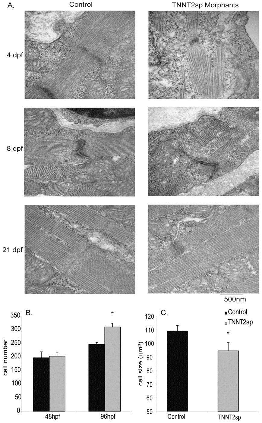
Fig. 3
Disruption of sarcomere structure and induction of myocardial hyperplasia in TNNT2sp morphants. (A) Representative electron micrographs of 96 hpf (top), 8 dpf (middle) and 21 dpf (bottom) ventricular cardiomyocytes. (B) Total cardiomyocytes at 48 hpf and 96 hpf. (C) Cardiomyocyte area at 96 hpf. *P<0.05; n=3–5 hearts per time point. All data expressed as mean + s.e.m.
Figure Data
Acknowledgments
This image is the copyrighted work of the attributed author or publisher, and
ZFIN has permission only to display this image to its users.
Additional permissions should be obtained from the applicable author or publisher of the image.
Full text @ Dis. Model. Mech.