
Fig. S5
- ID
- ZDB-IMAGE-160913-19
- Publication
- Tuttle et al., 2014 - Rabconnectin-3a regulates vesicle endocytosis and canonical Wnt signaling in zebrafish neural crest migration
- All Figures
- Figures for Tuttle et al., 2014
|
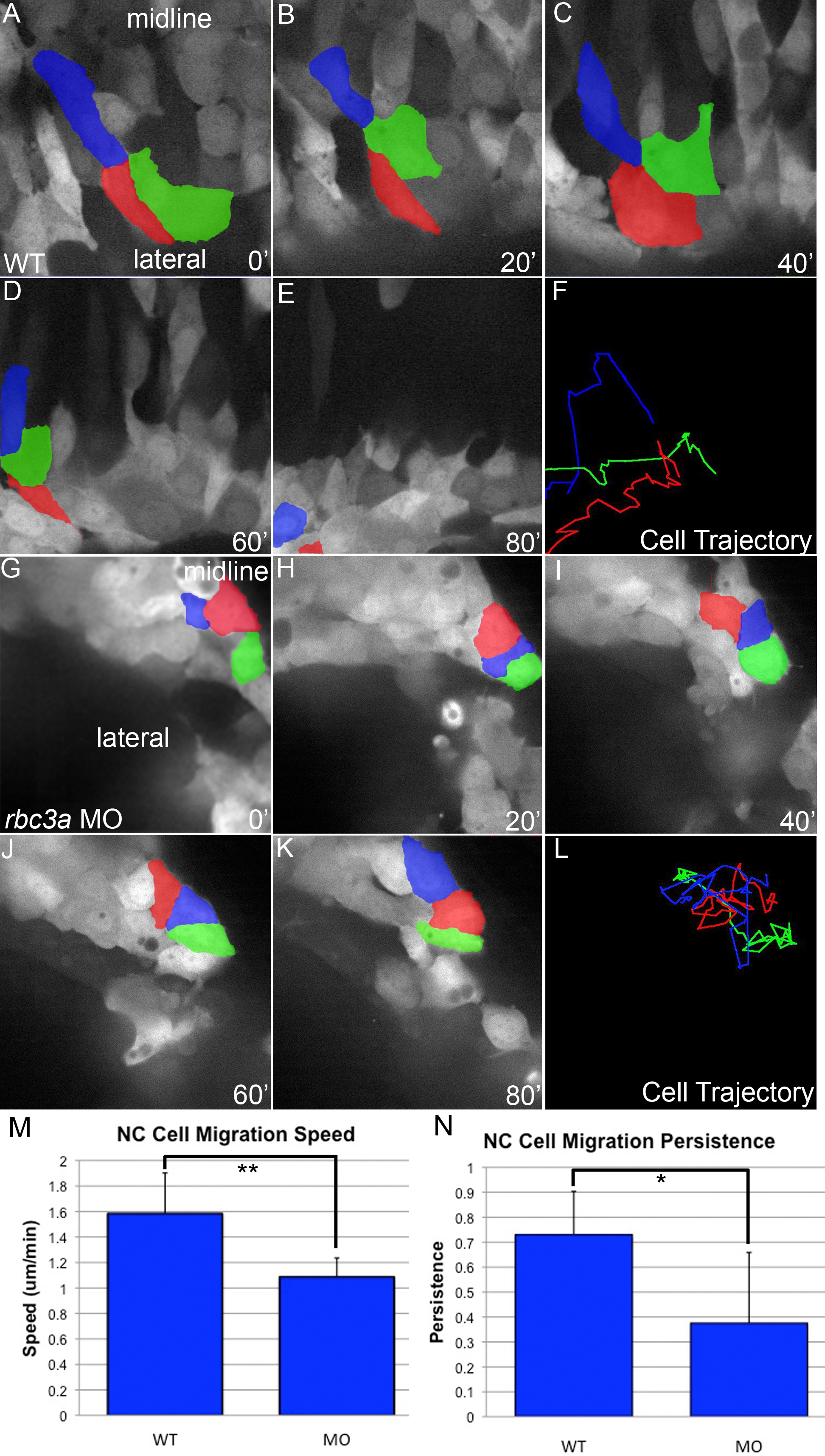
Fig. S5
Reduced NC cell motility in rbc3a-MO1-injected embryos. Individual frames from confocal time-lapsed movies of wild-type (A-E) and rbc3a-deficient (G-K) embryos from 13 hpf onwards in 20-min intervals. (F, L) Cell trajectories over 2 h of the corresponding cells in (A, F, and G-K). Wild-type NC cells (F) display stereotypical rapid, directed movement laterally and anteriorly, while many NC cells in rbc3a-MO1-injected embryos (L) adhere to each other and fail to migrate with other NC cells. (M) Average NC cell migration speed and (N) persistence of directionality (measured as the total displacement from the starting position of a cell over the total path length) at the onset of migration in wild-type and rbc3a-MO1-injected embryos (MO). Compared to wild-types, rbc3a-MO1 injection led to significantly reduced migration speed (p = 0.0028, 1.58±0.32 and 1.09±0.15 µm/min, respectively) and persistence (p = 0.015, 0.73±0.17 and 0.38±0.28, respectively). Error bars represent ±SEM. * p<0.05, ** p<0.01.