
IMAGE
Fig. 2
- ID
- ZDB-IMAGE-160909-2
- Publication
- Noonan et al., 2016 - Loss of vhl in the zebrafish pronephros recapitulates early stages of human clear cell renal cell carcinoma
- All Figures
- Figures for Noonan et al., 2016
Image
|
Figure Caption
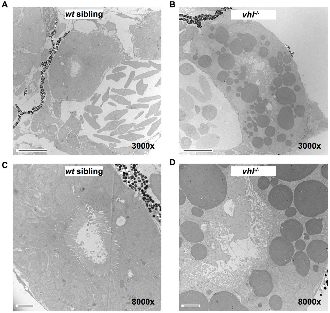
Fig. 2
TEM confirms that proximal vhl-/- pronephric tubules have an increased tubule diameter, a distorted lumen, disorganized cilia and contain cytoplasmic lipid vesicles. 5.5dpf vhl-/- and wt siblings were fixed, embedded and sectioned. Thin sections were stained with electron-dense metals and electron microscopy was performed. Transmission electron micrographs of proximal vhl-/- pronephros (B,D) in comparison to wt sibling (A,C). Scale bars: 10µm (A,B); 2µm (C,D).
Figure Data
Acknowledgments
This image is the copyrighted work of the attributed author or publisher, and
ZFIN has permission only to display this image to its users.
Additional permissions should be obtained from the applicable author or publisher of the image.
Full text @ Dis. Model. Mech.