
Fig. S1
- ID
- ZDB-IMAGE-160830-5
- Publication
- Just et al., 2016 - The mediator complex subunit Med10 regulates heart valve formation in zebrafish by controlling Tbx2b-mediated Has2 expression and cardiac jelly formation
- All Figures
- Figures for Just et al., 2016
|
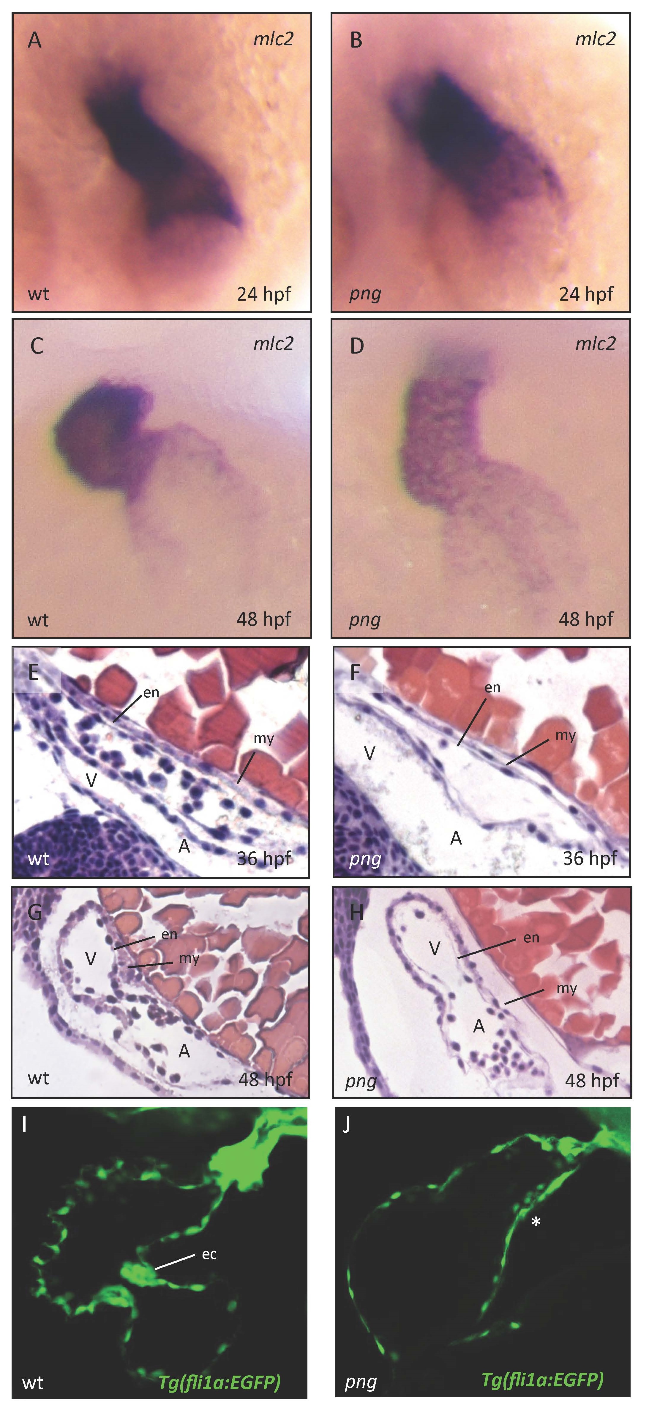
Fig. S1
Figure S1: Early cardiac development proceeds normally in png mutant embryos. Similar to wild-types (A, C), png mutant embryos (B, D) show a normal distribution of cmlc2 RNA at 24 and 48 hpf as revealed by whole-mount antisense RNA in situ hybridization. The heart regularly jogs to the left and loops to the right in wild-type (A, C) and png mutant (B, D) zebrafish embryos. (E-H) Hematoxylin/Eosin staining of sagittal histological sections of wt (E, G) and png mutant (F, H) hearts at 36 and 48 hpf, respectively. Similar to the wild-type situation (E, G), png mutant hearts (F, H) consist of normally developed endo- and myocardial cell layers. (I, J) Confocal images of Tg(fli1a:EGFP) transgenic wt (I) and png mutant (J) hearts at 72 hpf, showing that endocardial cushion cells fail to cluster at the AVC in png mutant hearts (marked with *). A, atrium; V, ventricle; my, myocardium; en, endocardium; ec, endocardial cushions.