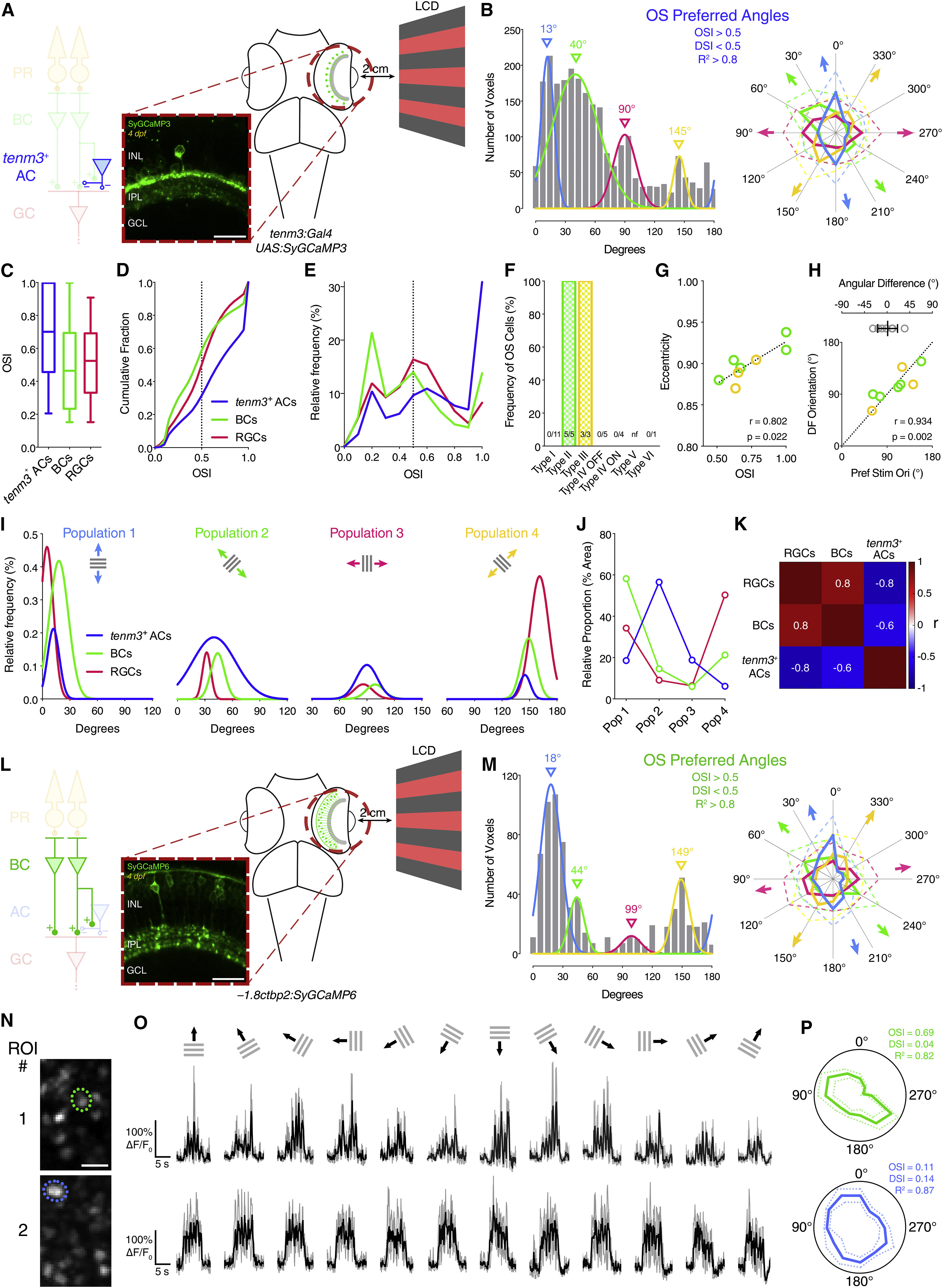
Fig. 6
Orientation Selectivity in Tenm3+ ACs and BC Terminals
(A) Two-photon functional calcium imaging of tenm3+ AC synaptic terminals expressing SyGCaMP3 (green) in 4-dpf Tg(tenm3:Gal4;UAS:SyGCaMP3) larvae. Distance of the eye from LCD screen is 2 cm. Recordings are performed from two to four Z-planes (approximately 20 µm total volume thickness). INL, inner nuclear layer; GCL, ganglion cell layer; IPL, inner plexiform layer. Scale bar, 20 µm.
(B) Cumulative histogram summarizing the incidence of preferred angles for identified tenm3+ AC OS voxels in 4-dpf larvae (n = 20 larvae). Overlaid curves are the fitted Gaussian distributions for each OS subtype. Polar plots illustrate the mean (+1 SD) normalized response profiles for each OS subtype.
(C-E) Degree of orientation selectivity (quantified by the OSI) across voxels with DSI < 0.5 and R2 > 0 in tenm3+ ACs (blue, n = 20 larvae), BCs (green, n = 20 larvae), and RGCs (red, n = 23 larvae; data from Figure 1H). Boxplots in (C) indicate interquartile ranges (boxes), medians (lines in boxes), and 10-90 percentiles (whiskers). The black dotted lines in (D) and (E) indicate the OSI threshold used to identify OS responses (OSI > 0.5).
(F) Bar histogram summarizing the frequency of OS cells among tenm3+ ACs in 4-dpf Tg(tenm3:Gal4) larvae injected with UAS:GCaMP6f DNA constructs (n = 29 cells from 27 larvae). The number of observed OS cells for each tenm3+ AC type is reported at the bottom. nf, not found.
(G) Scatterplot representing the relationship between OSI and dendritic field eccentricity of OS type II and III tenm3+ ACs (II, n = 5 cells; III, n = 3 cell). Spearman’s correlation coefficient (r) with the corresponding p value is reported. Dotted line represents the linear regression fit to the data.
(H) Scatterplot representing the relationship between preferred stimulus orientation and dendritic field orientation of OS type II and III tenm3+ ACs (II, n = 5 cells; III, n = 3 cell). Spearman’s correlation coefficient (r) with the corresponding p value is reported. Dotted reference line indicates x = y. Top graph shows the angular difference between preferred stimulus orientation and dendritic field orientation (mean ± SD).
(I) Normalized frequency distributions of preferred stimulus orientations in OS tenm3+ ACs (blue, n = 20 larvae), BCs (green, n = 20 larvae), and RGCs (red, n = 23 larvae; data from Figure 1J). The Gaussian distributions of the four different OS subpopulations are reported in separate graphs.
(J) Relative proportions of the four different OS subpopulations (Pop 1-4) in tenm3+ ACs (blue), BCs (green), and RGCs (red). Values are obtained by calculating the relative proportion (%) of the area under the normalized Gaussian curves in (I).
(K) Correlation matrix showing Spearman’s correlation coefficients (r) between the frequency distribution of the four OS subpopulations in tenm3+ ACs, BCs, and RGCs.
(L) Two-photon functional calcium imaging of BC ribbon synapses expressing SyGCaMP6 (green) in 4-dpf Tg(-1.8ctbp2:SyGCaMP6) larvae. Distance of the eye from LCD screen is 2 cm. Recordings are performed from two to four Z-planes (approximately 20 µm total volume thickness). Scale bar, 20 µm.
(M) Cumulative histogram summarizing the incidence of preferred angles for identified BC OS voxels in 4-dpf larvae (n = 20 larvae). Overlaid curves are the fitted Gaussian distributions for each OS subtype. Polar plots illustrate the mean (+1 SD) normalized response profiles for each OS subtype.
(N-P) Examples of visual responses to moving gratings from two BC terminals. Images showing the mean fluorescence across tuning experiments with identified regions of interest (ROIs) are reported in (N). (O) shows calcium responses of the two ROIs in (N) with black traces representing the average responses across three trials (gray traces) for each stimulus epoch. Polar plots in (P) illustrate the mean response profile (±SD, dotted lines) of each ROI with corresponding OSI, DSI, and R2 values. Note that ROI #1 shows orientation selectivity. Scale bar, 5 µm.
See also Figures S6 and S7, Table S1, and Movies S4 and S5.

