
Fig. 5
- ID
- ZDB-IMAGE-160824-5
- Publication
- Fontenille et al., 2014 - Microtubule-associated protein 9 (Map9/Asap) is required for the early steps of zebrafish development
- All Figures
- Figures for Fontenille et al., 2014
|
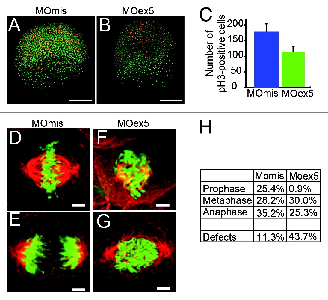
Fig. 5
Morpholino-mediated depletion of map9 leads to a reduction in the number of mitotic cells and to mitotic defects. (A and B) Embryos were injected at the 1-cell stage with 1 pmol MOex5 or MOmis (control) and stained with the anti-phosphorylated histone H3 (pH3) antibody (red). DNA was labeled with Hoechst 33258 (blue). Scale bars, 200 µm. (C) pH3-positive mitotic cells were counted in injected embryos at 5 hpf (n = 10/group). In MOex5 morphants the number of mitotic cells was reduced by ~40% in comparison to controls. Similar results were obtained in 8 hpf embryos (not shown). (D-G) Embryos were injected at the 1-cell stage with 1 pmol MOmis (control) (D and E) or MOex5 (F and G) and mitotic cells were imaged in 8 hpf embryos by confocal microscopy (scale bars, 10 µm). DNA was labeled with Hoechst (green) and MTs of the mitotic spindles with an anti-α-tubulin antibody (red). (H) Percentage of cells at different phases of mitosis. Mitotic cells were scored in map9 morphants and control embryos (n = 10 embryos/assay).