
Fig. 3
- ID
- ZDB-IMAGE-160823-25
- Publication
- Vihtelic et al., 2005 - Lens opacity and photoreceptor degeneration in the zebrafish lens opaque mutant
- All Figures
- Figures for Vihtelic et al., 2005
|
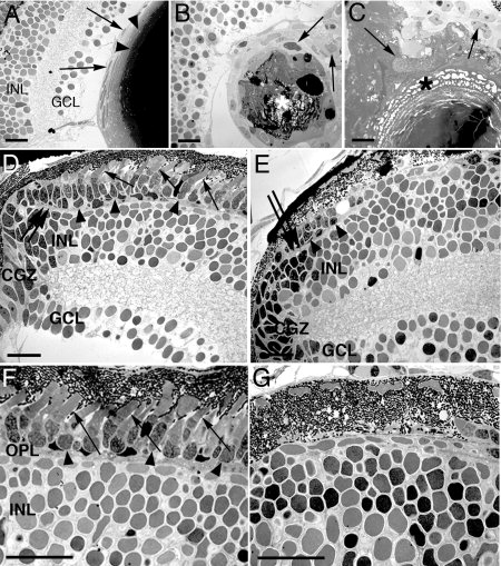
Fig. 3
Transmission electron microscopy of the lens and retina. A: The 7 days postfertilization (dpf) wild-type lens epithelial cells and underlying fiber cells are shown (arrows and arrowheads, respectively). B,C: Two different mutant lenses are shown. The mutant lenses display fiber degeneration in the lens nucleus and altered lens epithelial cell morphology (asterisks and arrows, respectively). D,F: The wild-type photoreceptor layer at the retinal margin and the central retina, respectively. The photoreceptor cell bodies and outer segments are labeled with arrowheads and arrows, respectively. E,G: In the mutant retina, a few remaining photoreceptor layer cell bodies are identified at the retinal margin (E, arrowheads), although no photoreceptor outer segments are evident. G: Similarly, the mutant central retina lacks identifiable photoreceptor cells. The outer plexiform layer in D and E is marked with a double arrow. INL, inner nuclear layer; GCL, ganglion cell layer; CGZ, circumferential germinal zone; OPL, outer plexiform layer. Scale bars = 20 µm in A-G.