
|
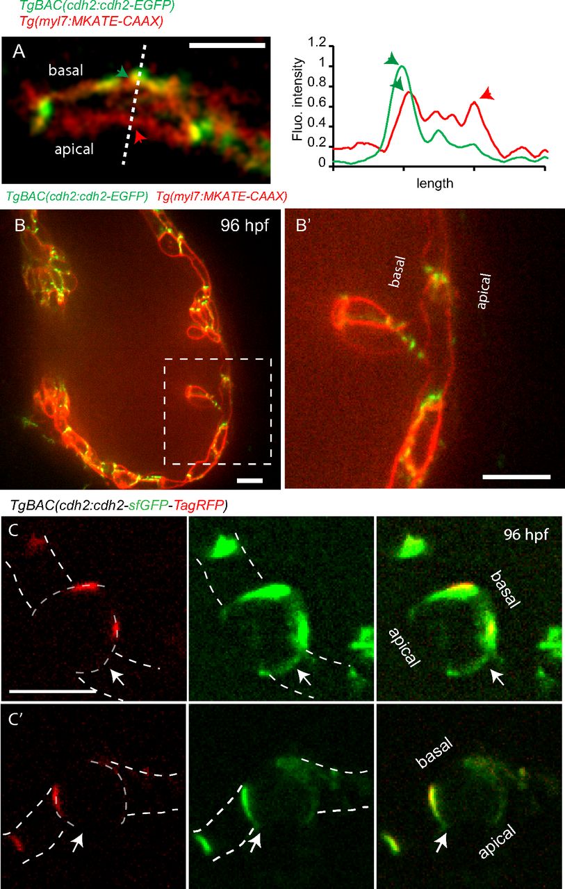
Fig. S5
Cdh2-EGFP molecules appear to move to the basal region of compact layer cardiomyocytes along the cell membrane. Spinning disk confocal images of a TgBAC(cdh2:cdh2-EGFP);Tg(myl7:MKATE-CAAX) heart. (A) Picture taken from time-lapse Movie S6; line scan quantification of Cdh2-EGFP and MKATE-CAAX; green and red arrows point to the basal and apical sides, respectively. (B′) Zoomed image of the region outlined by white dashed box. (B) Cdh2-EGFP is observed mainly on the cardiomyocyte’s cell membrane, and not in the cytoplasm. (C and C′) Spinning disk confocal images of TgBAC(cdh2:cdh2-sfGFP-TagRFP) cardiomyocytes. cdh2:Cdh2-tFT analysis indicates Cdh2 recruitment to the apical-lateral region of cardiomyocytes; white arrows point to a region with newly recruited Cdh2 displaying sfGFP signal without TagRFP signal. (Scale bar: 10 µm.)