
Image
|
Figure Caption
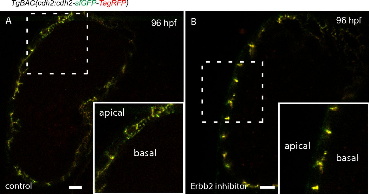
Fig. S10
Erbb2 signaling is required for Cdh2-EGFP distribution. (A) Red and green fluorescence sparsely distributed on the basal side of cardiomyocytes (control larva, left), indicating the dynamics of Cdh2-tFT. (B) Cdh2-tFT molecules are colocalized in cell-cell junctions, and thus show a yellow color in Erbb2 inhibitor-treated larva, suggesting the dynamics of Cdh2 are blocked. (Scale bars: 10 µm.)
Acknowledgments
This image is the copyrighted work of the attributed author or publisher, and
ZFIN has permission only to display this image to its users.
Additional permissions should be obtained from the applicable author or publisher of the image.
Full text @ Proc. Natl. Acad. Sci. USA