
Fig. 4
- ID
- ZDB-IMAGE-160810-23
- Genes
- Publication
- Sonal et al., 2014 - Myosin Vb Mediated Plasma Membrane Homeostasis Regulates Peridermal Cell Size and Maintains Tissue Homeostasis in the Zebrafish Epidermis
- All Figures
- Figures for Sonal et al., 2014
|
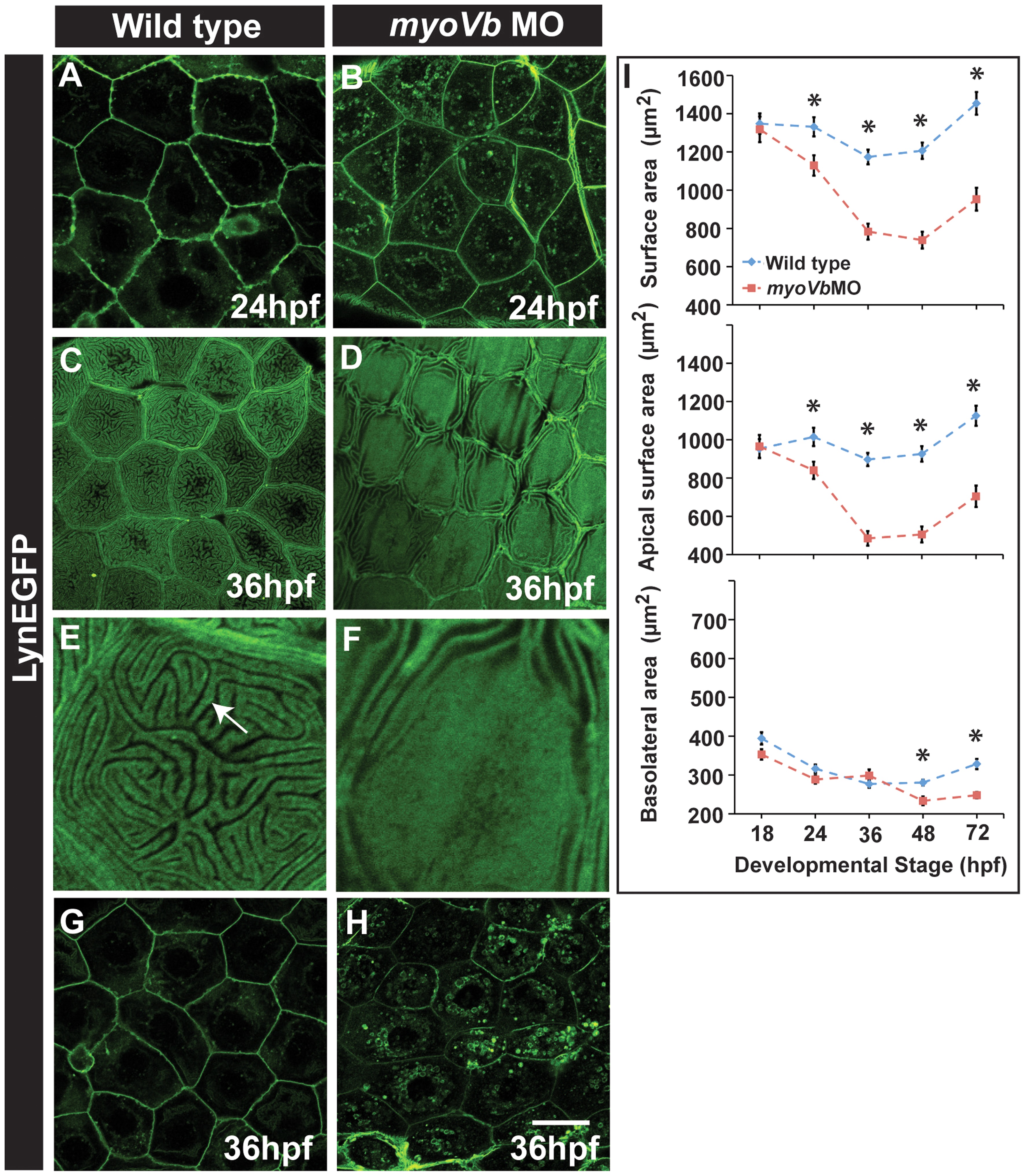
Fig. 4
Effect of loss of myosin Vb function on membrane projections, cell size and cell surface area.
LynEGFP staining in Tg(cldnB:lynEGFP) background revealed that membrane projections, which exist on basolateral side in wild type peridermal cells at 24hpf (A) are absent in myoVb morphants (B). Furthermore, apical microridges, present in wild type peridermal cells (C), are absent in myosin Vb morphants (D). E, F are digitally zoomed images of cells in ‘C’ and ‘D’, respectively. As compared to wild-type peridermal cells (G), morphant cells are smaller in size (H) at 36hpf. Quantification of cell surface area (I) reveals that total and apical surface area decreases substantially in morphants as compared to wild-type peridermal cells. The decrease in basolateral surface area is evident only after 48hpf. The error bars in ‘I’ represent standard errors of the mean whereas asterisks in ‘I’ indicate that the means are significantly different (student′s t test p≤0.05). Arrow indicates an apical microridge in a peridermal cell. Scale bars correspond to 20 µ in A-D and G,H.