
Fig. 3
- ID
- ZDB-IMAGE-160805-11
- Genes
- Antibodies
- Publication
- Wolman et al., 2015 - A Genome-wide Screen Identifies PAPP-AA-Mediated IGFR Signaling as a Novel Regulator of Habituation Learning
- All Figures
- Figures for Wolman et al., 2015
|
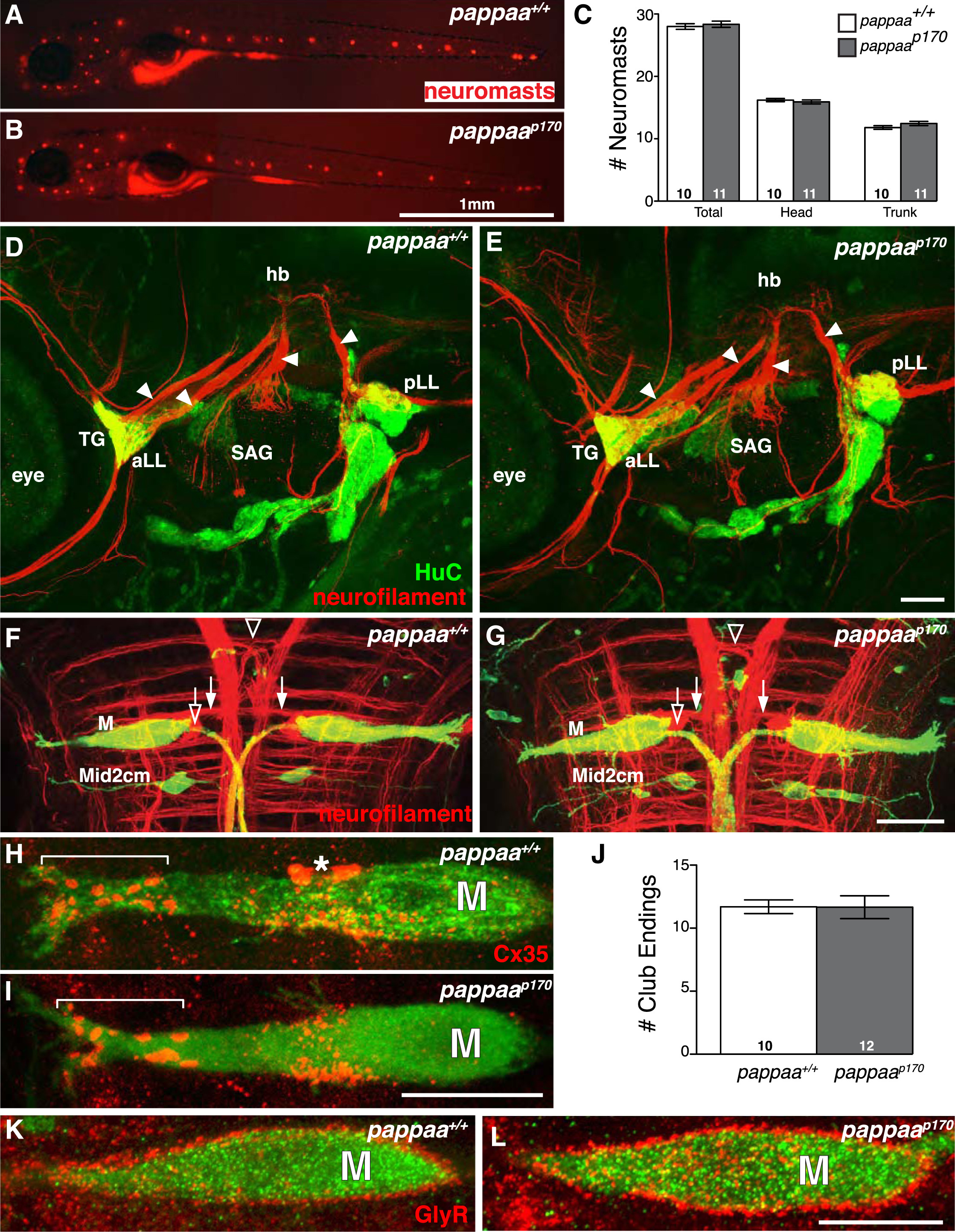
Fig. 3
Acoustic Startle Circuit Appears Intact in pappaap170 Larvae
(A-C) DASPEI labeling (A and B) and mean number (C) of sensory neuromasts.
(D-I and K-L) Projections of confocal stacks acquired at 5 dpf. Lateral views, anterior to the left in (A), (B), (D), and (E). Dorsal views, anterior to the top in (F)-(I) and (K)-(L). (D and E) Arrowheads mark projections (anti-neurofilament, red) from cranial ganglia (anti-HuC, green) to hindbrain. (F-I, K, L) Green label marks hspGFF130DMCA:UAS-gap43-citrine expression in Mauthner and Mid2cm Mauthner homologs. (F and G) Open arrows mark Mauthner (M) axon cap. Open arrowhead marks spiral fiber neuron projection and closed arrows mark contralateral passive hyperpolarizing (PHP) projection. (H and I) Brackets mark lateral dendrite of Mauthner with Cx35-positive club endings. Asterisk marks blood cell.
(J) Mean number of club endings.
(K and L) Glycineric receptors on Mauthner soma. SAG, statoacoustic ganglion; aLL, anterior lateral line ganglion; pLL, posterior lateral line ganglion. N, larvae shown within bars. Error bars indicate SEM. Scale bars = 1 mm (B), 50 µm (E), and 10 µm (G, I, and L).