
Fig. 2
- ID
- ZDB-IMAGE-160804-6
- Genes
- Antibodies
- Publication
- Rajaram et al., 2014 - Technical brief: Constant intense light exposure to lesion and initiate regeneration in normally pigmented zebrafish
- All Figures
- Figures for Rajaram et al., 2014
|
Fig. 2
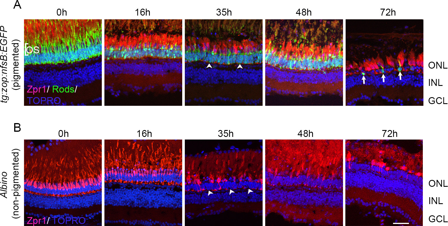
Photoreceptor loss in pigmented and nonpigmented retinas. Pigmented tg:zop:nfsB:EGFP zebrafish (A) or nonpigmented albino zebrafish (B) were subjected to intense light damage, and retinas were collected at the indicated time points and processed for immunohistochemistry. Retinas in (A) were stained with zpr-1 (red) and anti-green fluorescent protein (GFP; green) antibodies, while retinas in (B) were stained with zpr-1 (red) antibody; nuclei were counter-stained with TOPRO (blue). Dorsal retinas are shown. Arrowheads indicate regions of photoreceptor loss. Arrows denote rod photoreceptors that have rounded up and lost their outer segments. ONL, outer nuclear layer; INL, inner nuclear layer; GCL, ganglion cell layer; OS, outer segments. Scale bar is 50 µm.