
Fig. S4
- ID
- ZDB-IMAGE-160803-35
- Genes
- Antibodies
- Publication
- Shin et al., 2014 - Efficient homologous recombination-mediated genome engineering in zebrafish using TALE nucleases
- All Figures
- Figures for Shin et al., 2014
|
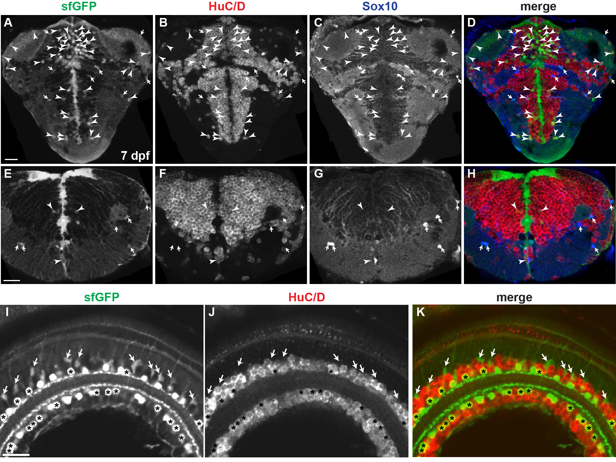
Fig. S4
Analysis of sfGFP-positive cells of sox2-2a-sfGFP line. Confocal microscope images. All images are transverse sections of 7 dpf sox2-2a-sfGFP larvae. (A-H) Arrowheads indicate HuC/D-positive, sfGFP-positive neurons. Arrows indicate Sox10-positive, sfGFP-positive OPCs. (A-D) Most sfGFP-positive cells are located in periventricular zones of the telencephalon and diencephalon. (E-H) Most sfGFP-positive cells are located in ventricular zones of the hindbrain. (I-K) Asterisks mark HuC/D-positive, sfGFP-positive cells that are putative amacrine cells. Arrows indicate HuC/D-negative, sfGFP-positive cells that are resembled as Müller glia morphologically.