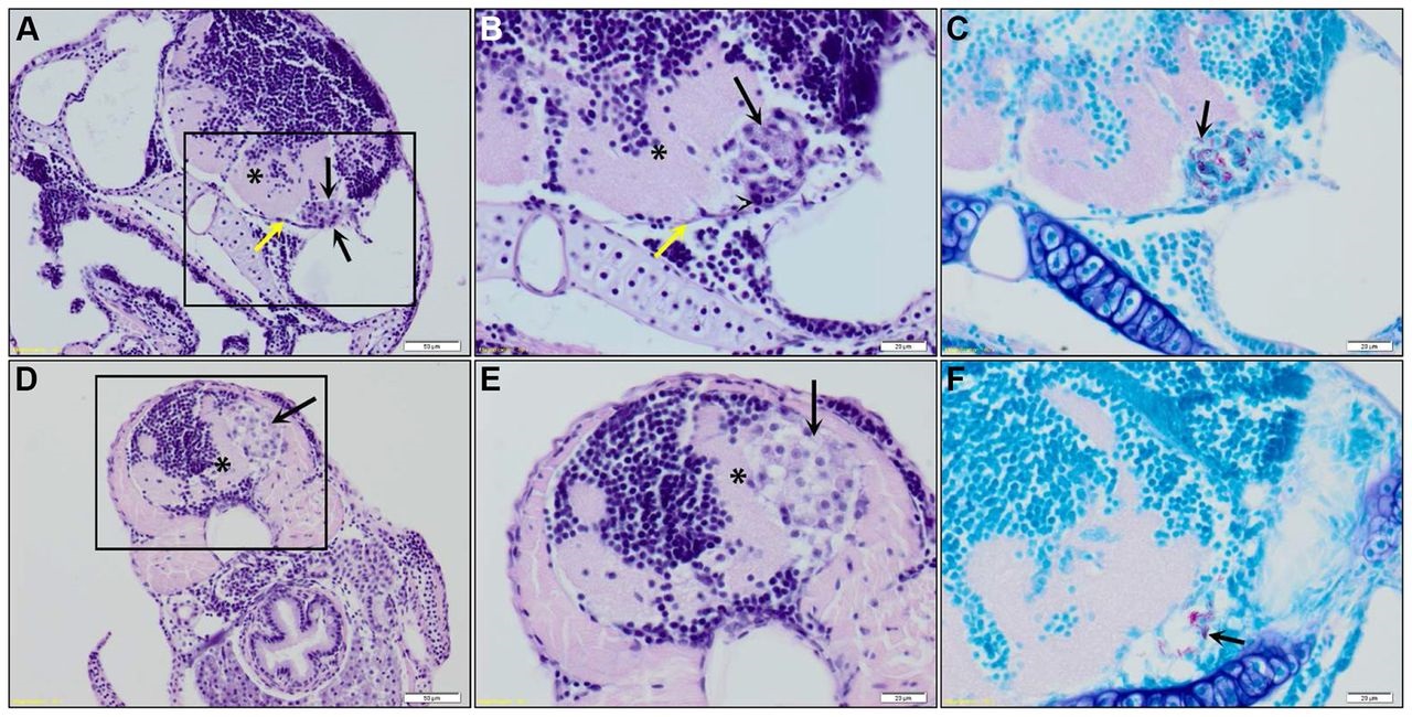
Fig. 3
Histopathological analysis of granulomas in the brain of a zebrafish embryo. (A) Coronal section of an embryo (7 dpf; 5 days after infection with M. marinum E11) showing an early granuloma (black arrows) attached to the meninges (yellow arrow), causing impingement of the underlying brain parenchyma (*). Scale bar: 50 µm. Marked area is enlarged in panels B and C. (B) The early granuloma is composed of epithelioid macrophages (black arrow) and heterophils (arrowhead). (C) ZN stain of granuloma depicted in panel B showing the presence of acid-fast bacilli (black arrow). Scale bars: 20 µm. (D) Coronal section of an embryo (7 dpf; 5 days after infection with M. marinum E11) showing an early granuloma (black arrow) present in the submeningeal space causing severe impingement of the underlying brain parenchyma (*). Scale bar: 50 µm. Marked area is enlarged in panels E and F. (E) The early granuloma is composed of foamy macrophages (black arrow) and (F) shows moderate numbers of acid-fast bacilli (black arrow). Scale bars: 20 µm.

