
Fig. 4
- ID
- ZDB-IMAGE-160727-49
- Genes
- Publication
- Hyatt et al., 1996 - Retinoic acid establishes ventral retinal characteristics
- All Figures
- Figures for Hyatt et al., 1996
|
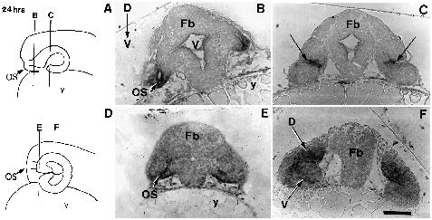
Fig. 4
The localization of pax[b] transcripts at 24 hpf in transverse sections (5 µm) of eyes from control embryos (B,C) and embryos treated with all-trans RA (E,F). (A) Schematic of a control embryo at 24 hpf indicating the level of transverse sections (lines B and C) shown in B and C, respectively. (B,C) In control embryos, transcripts are localized to the optic stalks (os in B) and to a sharp band extending across the retina (arrows in C) in the anterior region of the eyecup. (D) Schematic of an RA-treated embryo at 24 hpf illustrating the level of transverse sections (lines E and F) shown in E and F. (E,F) In treated embryos, pax[b] expression is present within the optic stalks (OS in E) and throughout the neuroepithelium of both the dorsal (D) and ventral (V) retina of the duplication. D-V, dorsal-ventral axis; y, yolk; bar, 65 µm in B, bar, 80 µm in C, E and F.