
Fig. 3
- ID
- ZDB-IMAGE-160727-48
- Genes
- Publication
- Hyatt et al., 1996 - Retinoic acid establishes ventral retinal characteristics
- All Figures
- Figures for Hyatt et al., 1996
|
Fig. 3
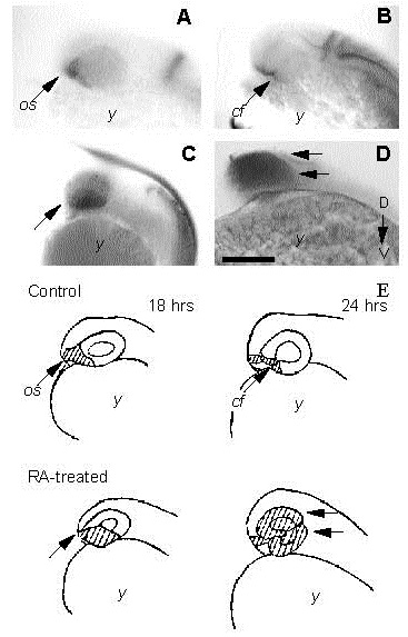
(A-D) A lateral view of control embryos (A,B) and RAtreated embryos (C,D) showing localization of pax[b] mRNA expression by whole-mount in situ hybridization. (A) An 18 hour control embryo with localization of pax[b] transcripts within the anterior/ventral region the eye adjacent to the optic stalk (os). (B) At 24 hpf, pax[b] transcripts in control embryos have been restricted to the optic stalk and within the choroid fissure (cf). (C) At 18 hpf, a ventral-to-dorsal spread of pax[b] transcripts is observed in RAtreated embryos. (D) By 24 hpf, pax[b] is expressed throughout the dorsal and ventral regions of the eyecups (double arrow) in treated embryos (arrow). (E) Diagrams of embryos shown above illustrates changes in pax[b] expression in control and RA-treated embryos. D-V, dorsal-ventral axis; y, yolk; bar, 210 µm.