
Fig. S5
- ID
- ZDB-IMAGE-160727-21
- Publication
- Kamaid et al., 2015 - Betaglycan knock-down causes embryonic angiogenesis defects in zebrafish
- All Figures
- Figures for Kamaid et al., 2015
|
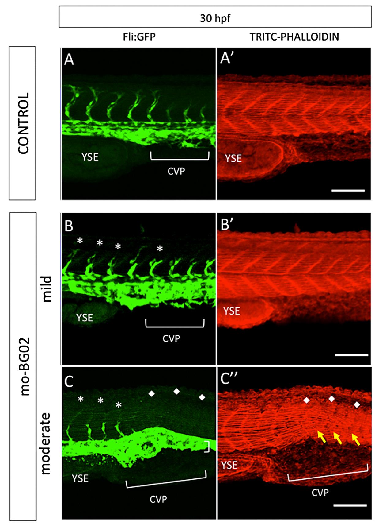
Fig. S5
zBG morpholino knockdown effects in caudal somite development. Images correspond to maximal projections of z-stack confocal images, showing the caudal somites of zebrafish Tg(fi1-eGFP)y1 embryos at 30 hpf. Representative examples of control embryos injected with 7 ng of mo-BG02-mis (A-A′), or morphant embryos injected with 7 ng of mo-BG02, and classified as mild (B-B′) or moderate (C-C′). GFP staining reveals endothelial cells in control (S5-A), mild morphants (S4-B), and moderate morphants (S5-C). A′-C′: TRITC-Phalloidin staining of the corresponding A-C images. In all pictures white asterisks (*) indicate incomplete or malformed Intersegmental vessels. White diamonds (♦) indicate total absence of ISVs in moderate morphants. Yellow arrows point to severely malformed somites in moderate morphants. Abbreviations: Intersegmental Vessel (ISV), Yolk Sack Extension (YSE), Caudal Vein Plexus (CVP)