
|
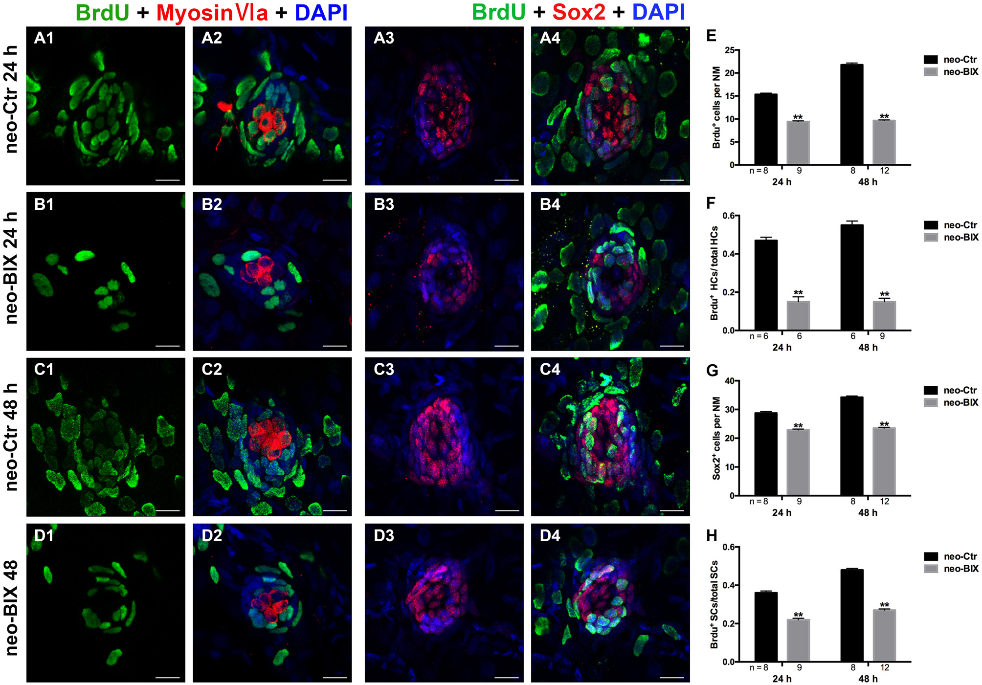
Fig. 5
BIX01294 significantly suppresses cell proliferation and induces reduced supporting cell (SC) production. We treated larvae at 5 dpf with neomycin and monitored cell proliferation with or without BIX01294 over the next 2 days. (A1-D2) The BrdU antibody shows dividing cells (green) in the NMs of the zebrafish lateral line, and HCs are stained with Myosin-VI (red). Scale bars = 10 µm. (A3-D4) Lateral line SCs are stained with Sox2 antibody (red), and nuclei are stained with DAPI (blue). The BrdU antibody shows dividing cells (green) in the NMs of zebrafish. Scale bars = 10 µm. (E) BrdU+ cells were counted in control and BIX01294-treated larvae at 24 h and 48 h after neomycin damage for 1 h. (F) Quantification of the ratio of BrdU+ HCs in control and inhibitor-treated larvae at 24 h and 48 h after neomycin incubation for 1 h. (G,H) The number of SCs and quantification of the ratio of BrdU+ SCs in control and BIX01294-treated larvae at 24 h and 48 h after neomycin incubation for 1 h. Bars are mean ± SD, and n = total number of embryos. **p < 0.001.