
Fig. S2
- ID
- ZDB-IMAGE-160715-32
- Publication
- Wei et al., 2016 - RNA polymerase III component Rpc9 regulates hematopoietic stem and progenitor cell maintenance in zebrafish
- All Figures
- Figures for Wei et al., 2016
|
Fig. S2
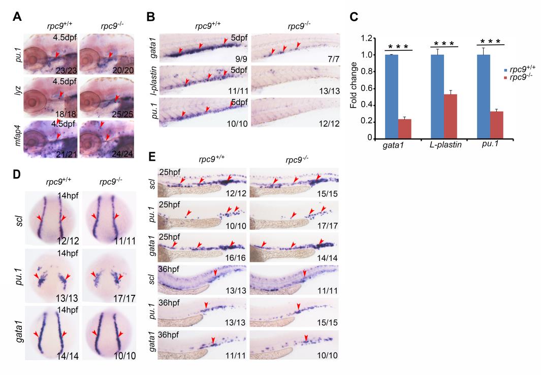
Hematopoietic cells are affected at 5 dpf but not at early stage in rpc9-/- embryos. (A) WISH result demonstrated the expression pattern of pu.1, lyz and mfap4 in the thymus of rpc9-/- or rpc9+/+ embryos at 4.5 dpf. Red arrowheads mark the thymus. (B) WISH result demonstrated that, compared to that of rpc9+/+ embryos, gata1, l-plastin and pu.1 were severely decreased in the CHT region of rpc9-/- embryos at 5 dpf. Arrowheads mark the CHT. (C) qRT-PCR result revealed that, compared with rpc9+/+ embryos, gata1, l-plastin and pu.1 were all significantly decreased in rpc9-/- embryos, at 5dpf. β-actin was used as internal control (mean±SD, ***P0.001). (D) WISH result demonstrated that, compared to that of rpc9+/+ embryos, scl, gata1 and pu.1 were not obviously altered in rpc9-/- embryos at 10 somite stage. Arrowheads mark the ALM(anterior lateral mesoderm) or PLM (posterior lateral mesoderm)region of zebrafish embryos. (E) WISH result demonstrated that, compared to the rpc9+/+ embryos, scl, gata1 and pu.1 in rpc9-/- embryos were not obviously altered at 25 and 36 hpf. Arrowheads mark the AGM or the CHT region.