
|
Fig. 2
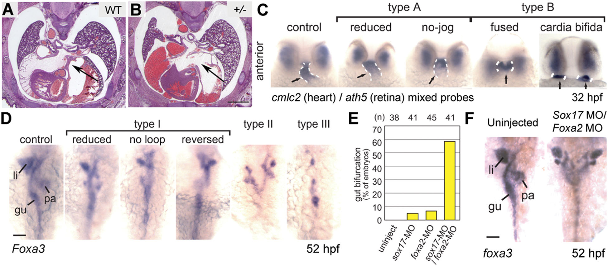
Heart and gut abnormalities in Nipbl-deficient animals. (A and B) Cryosections, stained with hematoxylin/eosin, of wildtype and Nipbl+/- littermate embryos at gestational day 17.5 (E17.5). A well-formed atrial septum is apparent in the wildtype heart, but absent in a significant fraction of mutants. Scale bar = 1 mm. (C) Zebrafish embryos injected with nipbla/b MOs show a range of defects in heart tube formation at 32 hpf, ranging from mild (type A) to severe (type B). Dashed white lines indicate heart tube, labeled by in situ hybridization (ISH) for a cmlc2 probe (ath5 ISH shows that eye is relatively unaffected). (D) Gut and visceral organ morphology assessed by ISH for foxa3 in nipbla/b morphants at 52 hpf. Defects ranged from mild looping defects (type I) to more severe defects such as bifurcation (type II) and even absence (type III) of gut parenchyma. gu, gut tube; li, liver; pa, pancreas. (E) Frequency of gut bifurcation in sox17 and foxa2 single and double morphants at 52hpf. (F) Example of severe gut bifurcation in sox17/foxa2 double morphant embryo. Scale bar in D and F = 50 µm. Adapted from [Kawauchi et al., 2009; Muto et al., 2011].