
|
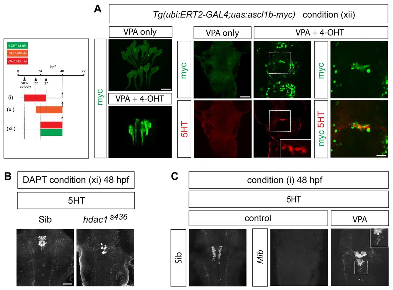
Fig. 5
Ascl1b is sufficient to rescue serotonergic differentiation in VPA-treated embryos. (A) Mis-expression of Myc-tagged Ascl1b in VPA-treated transgenic embryos rescues 5HT neuronal differentiation. A stable transgenic line expressing ERT2-GAL4 under the control of the ubiquitin (ubi) promoter was injected at the one-cell stage with plasmid DNA encoding Ascl1b-Myc under the control of UAS. Embryos were treated with VPA with or without 4-hydroxytamoxifen (4-OHT) [condition (xii)]. Myc immunostaining in shown in green. The upper and lower left panels show low-power views (scale bar: 500 µm) of Myc-immunostained zebrafish embryos treated with VPA in the absence (upper panel) or presence (lower panel) of 4-OHT. Addition of 4-OHT leads to widespread expression of Ascl1b-Myc (bottom left panel). Panels to the right show high-power views (scale bar: 50 µm) of Myc (green) and 5HT (red) immunostaining in the hindbrain of transgenic embryos. Embryos treated with VPA alone fail to express 5HT (n=51/51). Addition of 4-OHT and induction of Ascl1b-Myc expression rescues 5HT expression in 8/51 VPA-treated embryos (P=0.0058). Upper right and bottom right panels are high-power (scale bar: 20 µm) views of the boxed areas in the panels immediately to the left. The bottom, middle panel inset shows a higher-power view of the 5HT-expressing cells in the boxed area. The bottom right panel shows merged channels representing Ascl1b-Myc (green) and 5HT (red) immunostaining. (B) In hdac1s436 mutants treated with DAPT [condition (xi)], there is a rescue of 5HT neuronal differentiation at 48 hpf (n=22/22, P<0.0001). Scale bar: 50 µm. (C) On the mib mutant background there is a recovery of 5HT neurons at 48 hpf in VPA-treated embryos [condition (i)] (n=12/12, P=0.0002). Inset shows a high-power image of the boxed region.