Image
Figure Caption
Fig. S1
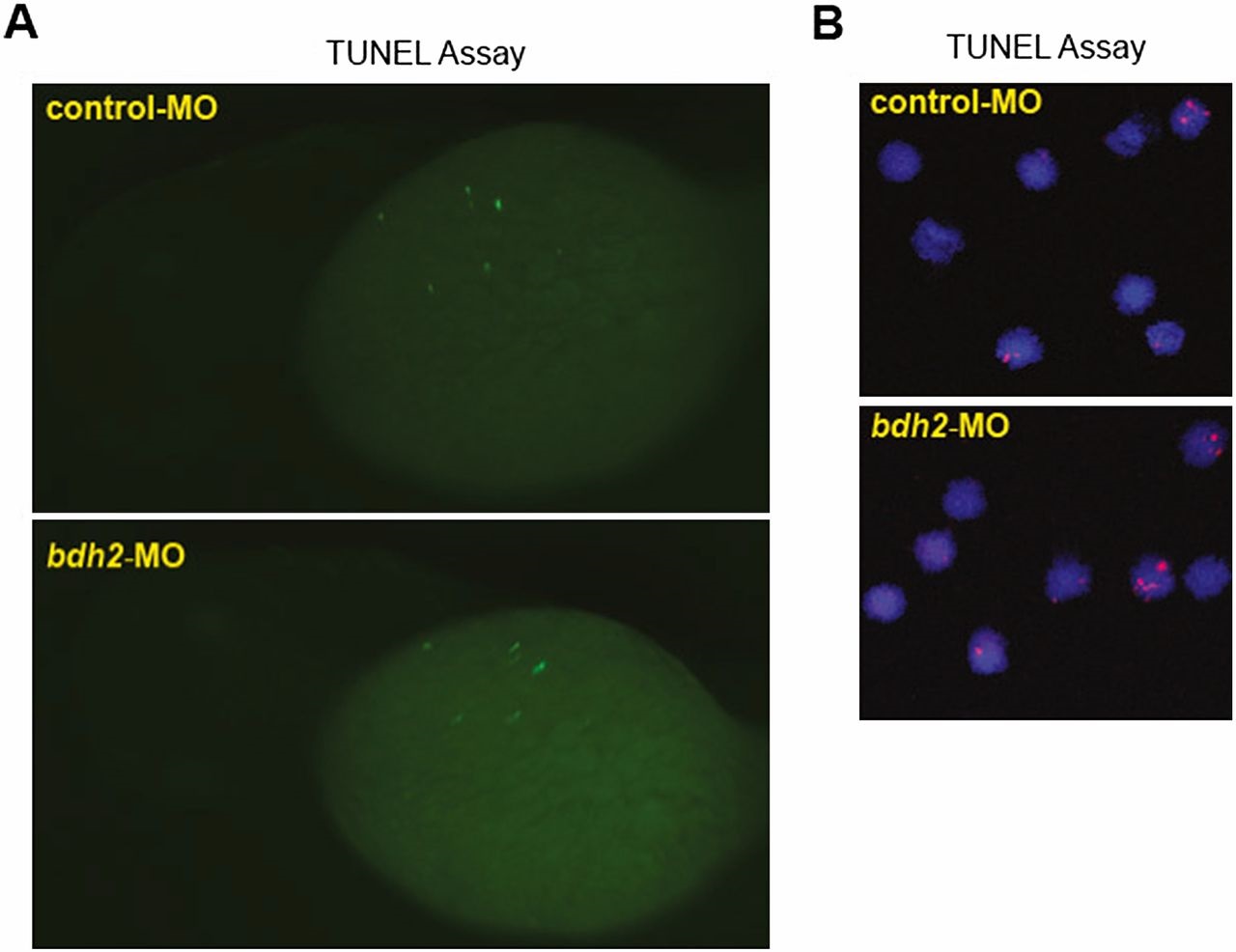
(A) TUNEL staining for apoptotic cells in control and bdh2 morphants. Control or bdh2 morphant embryos at 3 dpf were stained for apoptotic cells by using the TUNEL assay. Despite significant TUNEL staining in the nervous system, there is no excess staining of hematopoietic cells. Embryos are shown in lateral views. (B) TUNEL staining for apoptosis in erythrocytes isolated from control and bdh2 morphants at 3 dpf. Cells were counterstained with DAPI to visualize nuclei.
Acknowledgments
This image is the copyrighted work of the attributed author or publisher, and
ZFIN has permission only to display this image to its users.
Additional permissions should be obtained from the applicable author or publisher of the image.
Full text @ Proc. Natl. Acad. Sci. USA

